This site uses cookies to improve your experience. To help us insure we adhere to various privacy regulations, please select your country/region of residence. If you do not select a country, we will assume you are from the United States. Select your Cookie Settings or view our Privacy Policy and Terms of Use.
Cookie Settings
Cookies and similar technologies are used on this website for proper function of the website, for tracking performance analytics and for marketing purposes. We and some of our third-party providers may use cookie data for various purposes. Please review the cookie settings below and choose your preference.
Used for the proper function of the website
Used for monitoring website traffic and interactions
Cookie Settings
Cookies and similar technologies are used on this website for proper function of the website, for tracking performance analytics and for marketing purposes. We and some of our third-party providers may use cookie data for various purposes. Please review the cookie settings below and choose your preference.
Strictly Necessary: Used for the proper function of the website
Performance/Analytics: Used for monitoring website traffic and interactions
Ultrasound during cardiac arrest has quickly become standard. Initially, data suggested that the use of ultrasound during arrest increased pauses between compressions which worsens outcomes. Finally, patients with PEA and cardiac standstill on ultrasound have a 0.0%-0.6% Yours in ultrasounding, Shivam
Chuck Pilcher, MD, FACEP Editor, Medical Malpractice Insights Fibroids Found on Ultrasound Classic case of confirmation bias, appendicitis missed Facts : A middle-aged female with history of ovarian cysts and fibroids develops right lower quadrant abdominal pain rated as a 10 out of 10. To opt in to the free subscriber list, click here.
Casey Parker and James Rippey Ultrasound Case 112 A 30 year old woman who is currently 30 weeks gestation presents to the ED with abrupt, severe right loin pain.
Background Ultrasound is now readily available in the prehospital setting and its use has been highlighted as one of the top research priorities in prehospital care. This review evaluates the accuracy of prehospital ultrasound for the diagnoses of pneumothorax, haemothorax and pulmonary contusions in patients with trauma.
A point-of-care ultrasound (POCUS) was performed to evaluate for the presence of an FB using a high frequency (6–13 MHz) linear array ultrasound (US) transducer. There is a small 2–3 mm wound inferolateral to the right knee with very mild associated erythema, no active bleeding or swelling and no palpable FB ( figure 2 ).
Researchers at the University of California San Diego have created a wearable ultrasound system that can monitor deep tissues, as far as 16.5 Ultrasound-enabled wearables are enjoying a moment, with a variety of these technologies emerging recently. inches) below the surface of the body.
Background: The increased utility and accessibility of point-of-care ultrasound (POCUS) has allowed clinicians the freedom to rethink their diagnostic approach for many common diseases, including peritonsillar abscess (PTA). Test characteristics of ultrasound for the diagnosis of peritonsillar abscess: A systematic review and meta-analysis.
Casey currently splits his time […] The post SGEM#415: Buckle Down for some Ultrasound to Diagnosis Distal Forearm Fractures first appeared on The Skeptics Guide to Emergency Medicine. or there is a portable bedside ultrasound machine in the next room ready to go. He is also a fully-fledged ultrasonographer. Reference: Snelling et al.
Also, read this guide on how to practice ultrasound guided intravenous lines Sabrina Rodera Zorita Once the gelatin is firm, remove the pipes and transfer the model to a new container with water Try it, it’s very simple and cheap. See how it works!
The innervation of the knee is complex, but much of its sensory innervation is supplied by the genicular nerves, which are easy targets for ultrasound-guided nerve blocks. Step 2 Place the ultrasound system contralateral to the affected knee so that you have a clear line of sight of the ultrasound screen when performing the block.
Read this tutorial on the use of point of care ultrasonography (POCUS) for pediatric lung ultrasound. Take the ALiEMU PEM POCUS: Pediatric Lung Ultrasound Quiz Module Goals List indications for performing a pediatric lung point-of-care ultrasound (POCUS). Identify anatomical landmarks on ultrasound (Figure 3, Video 1).
They advise that the chest ultrasound should be used alone with caution. References: Lung ultrasound underdiagnoses clinically significant pneumothorax. Bottom line: This is an interesting and relatively large study. However, it is at odds with a paper published in 2021 from George Washington University. false negative rate.
IMAGING WITH ULTRASOUND Peritonsillar abscess is one of the most common deep space infections of the head and neck contributing significantly to health care costs in the United States. The use of point of care ultrasound for diagnosis and guidance of treatment may be an adequate alternative to obtaining CT imaging. 2012;147(3):472-474.
Acute cholecystitis on ultrasound. Image by James Heilman, MD - Own work, CC BY-SA 3.0, [link] The diagnosis of cholecystitis can be made quickly and accurately in the emergency department using point-of-care ultrasound, according to a new meta-analysis.
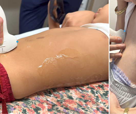
You set up everything, have the patient positioned, and then notice there is no sterile ultrasound gel. The trick is to eliminate anything of poor acoustic impedance between the ultrasound probe and the patient’s skin. Note that some ultrasound manufacturers do not recommend the use of isopropyl alcohol on their transducers.
Casey Parker and James Rippey Ultrasound Case 111 A 45 year old woman with chronic alcoholic liver disease presents to the ED with exertional dyspnoea and is noted to have a SpO2 of 90% at rest despite having a normal chest examination and CXR.
1 A biliary point-of-care ultrasound (POCUS) is the fastest and most accurate way to assess for biliary pathology, but it can be difficult to locate the gallbladder, as it is not a fixed organ. Ultrasound. Emergency physician-performed ultrasound to diagnose cholelithiasis: a systematic review. 4 Benefits of Biliary POCUS?
TheOttawa DVT PoCUS Handbookis a peer-reviewed, practical guide designed to support emergency clinicians in the bedside diagnosis of deep vein thrombosis using point-of-care ultrasound (PoCUS).
Researchers at the University of Connecticut have developed an ultrasound implant that can assist in opening the blood brain barrier to allow chemotherapy to enter and treat brain cancer. The researchers combined this with biodegradable polymers to create the ultrasound implant, which can be seen in the image above.
A bedside ultrasound is completed to assess the location of the pregnancy. A radiology performed ultrasound is ordered and has similar findings– Impression: no definitive IUP with a small amount of free fluid within physiologic limits. Laboratory evaluation reveals a hemoglobin and hematocrit of 12.6/37.1
Unlock the foundational skills you need to perform ultrasound-guided nerve blocks in this detailed tutorial! Courses The post Mastering Ultrasound-Guided Nerve Blocks Basics first appeared on Core Ultrasound.
Many thanks to Dr Manoj Wickramsinghe for his review of this fabulous POCUS textbook. He is a trainee in Anaesthesia and ICM, based in Leeds, and one of the CCN editorial team. About the book authors Editors; Hatem Soliman-Aboumarie, Marcelo Haertel Miglioranza, Luna Gargani, Giovanni Volpicelli. The authors and editors of this book include some.
Ultrasound diagnosis of occult pneumothorax. The role of point-of-care ultrasound in the diagnosis of pericardial effusion: a single academic center retrospective study. Ultrasound J. Pediatr Clin North Am. 2010 Dec;57(6):1221-34. PMID: 21111115. Crit Care Med. 2005 Jun;33(6):1231-8. PMID: 15942336. link] Hanson MG, Chan B.
St.Emlyn's - Emergency Medicine #FOAMed Part 1 of an introduction to paediatric ultrasound in the emergency department from Dr Pete Hulme @Dr_Pete_EmMed @stemlyns #FOAMed The post Paediatric Point of care ultrasound: Big Kids playing with toys or the future of Paediatric emergency medicine?
Take the ALiEMU PEM POCUS: Soft Tissue Quiz Case Goals List the indications of performing a pediatric soft tissue point-of-care ultrasound (POCUS). Pediatric Soft Tissue POCUS Ultrasound Technique Figure 1. Linear ultrasound transducer Probe Use a linear, high-frequency transducer. Describe the limitations of soft tissue POCUS.
St.Emlyn's - Emergency Medicine #FOAMed Part 2 of our introduction to ultrasound use in the paediatric emergency department with @Dr_Pete_EmMed #USS @FOAMed #paediatrics The post Paediatric Point of care ultrasound: Big Kids playing with toys or the future of Paediatric emergency medicine?
A bedside right upper quadrant ultrasound was performed, and the images are below. The presence of gallstones outside of the gallbladder wall, striated appearance of the gallbladder wall, and adjacent fluid collections, abscesses or fistulas are other findings that could be seen on ultrasound (5, 8). 2018;34(2):132-136.
Researchers at MIT have developed a wearable ultrasound system that is intended to allow women at high risk of breast cancer to perform an ultrasound scan on themselves at home, and may also let patients with early-stage malignancy or suspicious lesions to monitor how they are progressing.
For those new to the probe, we recommend first reviewing the basics in the incredible FOAMed Introduction to Bedside Ultrasound Book , 5 Minute Sono , and POCUS Atlas. Understanding the ultrasound image in relation to your probe is fundamental to any ultrasound procedure! Then holding the ultrasound still when moving the needle.
I know we already know this, but it especially true when you are talking about trying to obtain images on a wiggling fetus somewhere around the size of a banana.
Given her pain with a history of intermittent hematuria and dysuria, you perform a renal and bladder point of care ultrasound (POCUS) examination. Pre-warmed ultrasound gel is helpful when available. Then test your skills on the ALiEMU course page to receive your PEM POCUS badge worth 2 hours of ALiEMU course credit.
Ultrasound can assist: confirm ascites, evaluate for best site, abdominal wall thickness, blood vessels along needle track. Safety of ultrasound-guided thoracentesis in patients with abnormal preprocedural coagulation parameters. Paracentesis is a safe procedure with a low complication rate (< 1%). 2013;144:456–463. Am J Emerg Med.
Kenny MD [ @heart_lung ] Since its first description [1], the Venous Excess Ultrasound Score [VExUS] has received much research attention. Subsequently in the late 1970s, Sivaciyan and Ranganathan [9] confirmed these findings with Doppler ultrasound of the IJV. The ultrasound journal 12: 1-12 2. Jon-Emile S. Echocardiography 7.
Ever been in a trauma activation where it seems like the first thing that happens is that someone steps up to the patient with the ultrasound probe in hand? Then pull out the ultrasound machine, but be quick about it. And then it takes 5 minutes of pushing and prodding to get the exam done? Well, it’s not supposed to be that way.
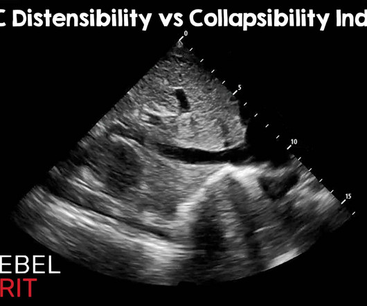
Regarding caval indexes, the advent of artificial intelligence and advanced learning has become integrated into many ultrasound machines. Ultrasound Med Biol. As with most things in medicine, it is important to understand the pitfalls. Most measurements are somewhere around the hepatic confluence. May 2014; PMID: 24495437.
Background: Point-of-care ultrasound (PoCUS) is a valuable clinical tool in the assessment of acute dyspnea. Impact of serial cardiopulmonary point-of-care ultrasound exams in patients with acute dyspnoea: a randomized, controlled trial. PoCUS evaluations included lung ultrasound (LUS) and focused cardiac ultrasound (FoCUS).
Ocular point-of-care-ultrasound (POCUS) was performed as seen below. Our patient underwent anterior orbitotomy, but there are case reports in the literature of ultrasound-guided drainage (4). In addition, there are studies that discuss missed cases of abscess on CT that were detected with use of ultrasound. Brzycki et al.
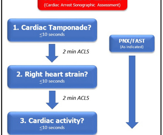
In part two of this series on using ultrasound during cardiac arrest, we dive into advanced strategies to further optimize your resuscitation care. Building on the foundation from part one, this video focuses on actionable tips to take your ultrasound skills to the next level during cardiac arrest scenarios.
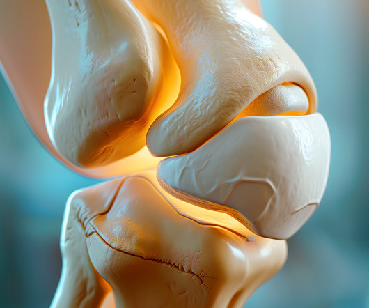
Eckler, MD discuss the March 2025 Emergency Medicine Practice article, Emergency Department Management of Knee Pain Common Etiologies of Knee Pain Risk Factors and Statistics Infectious Causes of Knee Pain Pre-Hospital Care and EMS History and Physical Exam Imaging Guidelines Ottawa Knee Rule and X-Ray Necessity Imaging Modalities for Knee Effusion (..)
Ultrasound is a powerful tool that can significantly enhance patient care during cardiac arrest. In Part 1 of this lecture, we break down how ultrasound can be effectively utilized in three key areas: identifying reversible causes, assisting in critical procedures, and guiding resuscitation.
A flurry of tests later, my ultrasound showed good doppler flow, and my lab work was pristine. I was in the OR for the first day of my anesthesia rotation when suddenly the pain hit me. It was in my left flank, radiating to my back — so much pain I could hardly think.
University of Maryland Department of Emergency Med
NOVEMBER 19, 2023
Acute bronchiolitis (AB) is a common cause of respiratory tract infections in infants. A recent study looked at the application of Point-of-Car. Click to view the rest
We organize all of the trending information in your field so you don't have to. Join 5,000+ users and stay up to date on the latest articles your peers are reading.
You know about us, now we want to get to know you!
Let's personalize your content
Let's get even more personalized
We recognize your account from another site in our network, please click 'Send Email' below to continue with verifying your account and setting a password.


Let's personalize your content