EM@3AM: Brainstem Strokes
EMDocs
MAY 11, 2024
Answer : Brainstem stroke specifically in the pons resulting in locked in syndrome. CT head without contrast 1 is performed and reveals the following: Question: What is the diagnosis?
This site uses cookies to improve your experience. To help us insure we adhere to various privacy regulations, please select your country/region of residence. If you do not select a country, we will assume you are from the United States. Select your Cookie Settings or view our Privacy Policy and Terms of Use.
Cookies and similar technologies are used on this website for proper function of the website, for tracking performance analytics and for marketing purposes. We and some of our third-party providers may use cookie data for various purposes. Please review the cookie settings below and choose your preference.
Used for the proper function of the website
Used for monitoring website traffic and interactions
Cookies and similar technologies are used on this website for proper function of the website, for tracking performance analytics and for marketing purposes. We and some of our third-party providers may use cookie data for various purposes. Please review the cookie settings below and choose your preference.
RebelEM
FEBRUARY 24, 2025
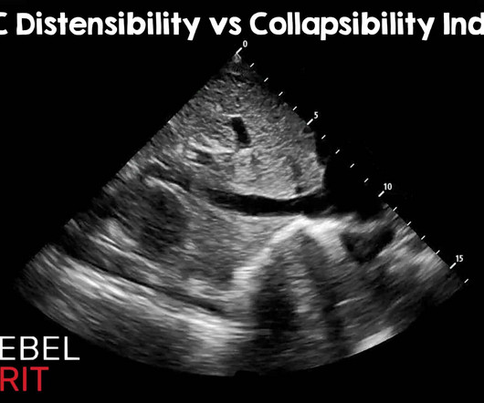
Of course, there are other methods of assessing fluid tolerance : Capillary refill evaluation, passive leg raise, central venous pressure measurement, pulmonary artery wedge pressures, stroke volume variation, pulse pressure variation, etc. Ultrasound Med Biol. Most measurements are somewhere around the hepatic confluence.
This site is protected by reCAPTCHA and the Google Privacy Policy and Terms of Service apply.
Sensible Medicine
NOVEMBER 6, 2023
Many doctors believe that closing the left atrial appendage (with a device) will help reduce stroke and bleeding. The idea behind stroke reduction is that occluding the appendage takes away a common area where clots form. I’ve already told you that peri-device leaks are associated with an increase in stroke risk.
EMDocs
MARCH 19, 2024
Thromboembolism can lead to stroke (and this is a more common cause of stroke in the setting of sCAD than hypoperfusion). Epidemiology Common cause of strokes in young people; sCAD accounts for 15-24% of strokes in patients < 45 years. Rare cause of stroke overall – incidence is 1.72
Pediatric Education
APRIL 30, 2023
She had a history of neonatal stroke for which she had received physical and occupational therapy. The mother and medical records confirmed that a cause had not been determined but the child had a neonatal arterial ischemic stroke. The mother had been evaluated for hypercoagulability which was negative.
Sensible Medicine
DECEMBER 28, 2023
The idea is to prevent stroke and reduce bleeding by plugging the appendage. But, get this: PREVAIL did not find noninferiority in its first co-primary endpoint of stroke, systemic embolism or cardiovascular death. One makes an ultrasound device, the other a radiofrequency device. This was a recent FDA approval.
Sensible Medicine
JUNE 28, 2024
She is admitted with suspected stroke. Case 1: Excess An elderly woman is admitted to a community hospital with a minor stroke. And then, I guess because cancer-related hypercoagulability could conceivably cause a stroke, they went further. Sensible Medicine is a reader-supported publication. I love my job.
Mind The Bleep
FEBRUARY 2, 2025
For example: “If ultrasound negative then reassure and discharge”, “If CRP normal then safety net and advise to see GP if symptoms recur” Treatment Escalation Plan/ DNACPR Clearly document if the patient has an existing DNACPR in place- they may come to hospital with a copy.
Sensible Medicine
JUNE 17, 2024
Stroke occurred in 7 patients (0.12%). Notable also that in the main paper, there was one TIA in the PFA arm and one stroke in the thermal arm but neither of these were in the MRI subgroup of the study. We really can’t say much about safety when only 600 patients are randomized. We would call these silent brain lesions.
PulmCCM
NOVEMBER 17, 2023
For any given hemodynamic state, measure or estimate the P ra and CO [or stroke volume, SV] simultaneously and repeatedly after each intervention. ’ This could be done with the Venous Excess Ultrasound Score [VExUS] [5, 6]. The ultrasound journal 2020, 12(1):1-12. Ultrasound J 2022, 14(1):36. What does this mean?
RebelEM
JANUARY 29, 2024
Randomized, Controlled Trial of Ultrasound-Assisted Catheter-Directed Thrombolysis for Acute Intermediate-Risk Pulmonary Embolism. A prospective, Single-Arm Multicenter Trial of Ultrasound-Facilitated, Catheter-Directed, Low-Dose Fibrinolysis for Acute Massive and Submassive Pulmonary Embolism: The SEATTLE II Study. CHEST 2010.
Taming the SRU
AUGUST 7, 2023
The patient needs a transvaginal ultrasound w/ doppler with gynecology consult and possible laparotomy TVUS is not just for torsion, it can also be used for ectopic pregnancies or pregnancy of unknown location Acute Ischemic Stroke Updates WITH Drs.
ALiEM
JANUARY 29, 2024
It requires immediate consultation with ophthalmology as well as neurology as it is considered a stroke equivalent. Stroke workup for CRAO is necessary, and don’t forget about secondary prevention/risk stratification which must be part of the management. RBSS may predict poor response to systemic thrombolysis. 39,1 (2020): 197-202.
Taming the SRU
NOVEMBER 3, 2023
Ultrasound‐Guided Serratus Anterior Plane Block (SAPB) Improves Pain Control in Patients With Rib Fractures. Ultrasound Med. 41, 2695–2701 (2022). Materials and Methods It was an interventional study at a single tertiary care referral center with a level I trauma designation and an approximate annual ED census of 90,000.
Taming the SRU
FEBRUARY 5, 2024
It is most helpful to do the ultrasound immediately before needle insertion, as movement of the patient may shift cutaneous landmarks from underlying bony structures. This resource offers additional information on ultrasound assisted LP’s. WHY - Why Not? REFERENCES 1. Zambito Marsala, S., Gioulis, M., & Pistacchi, M.
Dr. Smith's ECG Blog
APRIL 2, 2024
Bedside ED ultrasound showed exceedingly poor global LV function, and no B lines. No wall motion abnormality Decreased left ventricular systolic performance, severe Left ventricular enlargement, marked Dilated cardiomyopathy severe Est. stroke volume 52 cc at HR 70 = 3.64 Here is the initial ED ECG. What do you think?
EMDocs
MAY 15, 2024
Ultrasound Sensitivity 88-100%, specificity 68-94% Positive likelihood ratio of 14.6 (95% While chest radiograph is often the first-line imaging modality, lung ultrasound has higher sensitivity and can detect pulmonary abnormalities earlier; CT imaging should be considered if initial imaging is negative or if concern for PE is present.
ACEP Now
OCTOBER 29, 2024
Passively, I watch another clinician’s cursory examination of a critically ill patient to determine how they deem a patient “toxic-appearing” or their bedside neurological assessment to activate a code stroke. A testicular ultrasound confirmed restored blood flow. What do you think the prognosis is for that patient we achieved ROSC?
Dr. Smith's ECG Blog
JULY 5, 2024
But the duration of symptoms was prolonged and so to avoid the risk of stroke, AV nodal blockade (rate control) was preferred. We want to avoid a stroke. A formal ultrasound later showed reasonably good LV function, and so he later received carvedilol and diltiazem, Unfortunately, those led to hypotension at 80/40 with a HR 40.
RebelEM
MARCH 8, 2023
CRAO is a stroke of the eye; patients should be considered for a complete stroke work up. Signs Shafer’s sign (tobacco dust) May see a Weiss ring when the posterior vitreous (PV) detaches from the optic disc margin Visualization with ultrasound. Emergency Department Management: Emergency ophthalmology consultation. Read more
EMDocs
MAY 28, 2024
Ultrasound Sensitivity 88-100%, specificity 68-94% LR+ of 14.6 (95% While chest radiograph is often the first-line imaging modality, lung ultrasound has higher sensitivity and can detect pulmonary abnormalities earlier; CT imaging should be considered if initial imaging is negative or if concern for PE is present. pneumoniae, C.
RebelEM
JUNE 26, 2023
Severe acute traumatic coagulopathy = PT >1.5 to 3.33; P = 0.72 NOT STATISTICALLY SIGNIFICANT Also no difference in individual components given Thromboembolic Events: 4F-PCC: 35% Placebo: 24% Absolute Diff: 11%; 95% CI 1 to 21% Relative Risk 1.48; 95% CI 1.04 to 2.10; P = 0.03 Severe acute traumatic coagulopathy = PT >1.5 to 3.33; P = 0.72
RebelEM
JUNE 19, 2023
SBP < 75 mm Hg: 39% SBP 76-89 mm Hg: 33% Critical Findings: No statistically significant difference in safety outcomes (DVT, PE, MI, Stroke) TXA Placebo RR (95% CI) Primary Outcome Good Functional Outcome (6 months) 53.7% Patients were actively screened for DVT (all received lower extremity ultrasound on or around day 7).
PulmCCM
JULY 10, 2023
Read in JAMA When to start anticoagulation after ischemic stroke from atrial fibrillation It’s been unclear when to start anticoagulation after an ischemic stroke presumed due to thromboembolism from atrial fibrillation. Aspirin is sometimes given during the waiting period. There were two intracranial hemorrhages in each group.
ACEP Now
JUNE 6, 2023
Notoriously elusive, with a high misdiagnosis rate, thoracic aortic dissection (AD) can mimic many conditions, including acute coronary syndrome (ACS, the most common), gastroesophageal reflux disease (GERD), stroke, and spinal-cord compression. 1 Opioids or anxiolytics are often given to patients whose diagnosis of AD is missed or delayed.
EM Curious
SEPTEMBER 9, 2015
CUTTING EDGE ACUTE ISCHEMIC STROKE CARE [LECTURE]: Dr. Melissa Motta, a neuro-critical care expert breaks down current best practice in acute ischemic stroke in an excellent talk at Maryland CC Project. SEE YOU NEXT WEEK Online Advanced Ultrasound Education Northwestern EM Residency Education Coming soon.
Resus Review
OCTOBER 9, 2015
However, advancements in percutaneous coronary intervention (such as drug-eluting stents and techniques, risk stratification, and intravascular ultrasound) have increased the viability of this treatment option. The development of DES stents and second generation antiplatelet medications and reduced the incidence of in stent restenosis.
Northwestern EM Blog
MAY 16, 2022
What is your initial imaging test of choice, ultrasound (US) or non-contrast CT, and why? Would you be confident in a point-of-care-ultrasound evaluation or a formal ultrasound? Many patients in the ultrasound groups did get additional imaging, but this was not the majority. How do you proceed? In this study, 40.7%
Taming the SRU
FEBRUARY 24, 2024
Providers should use ultrasound as early as possible to evaluate for cardiac dysfunction, pulmonary edema, and fluid status. They tend to be hypertensive, tachycardic, tachypneic, and hypoxic. Do not always appear volume overloaded on exam. Early recognition and prompt treatment is key.
Taming the SRU
JUNE 3, 2024
Susan Wilcox Increasing regionalization due to: Growth of specialty centers Increasing development of healthcare systems Hub-and-spoke models Development of ECMO, trauma, transplant, and stroke centers The higher the acuity of the centers, the higher the acuity of the patient that needs to get there.
Taming the SRU
AUGUST 4, 2024
In select patient populations (such as those with poor outpatient follow-up), screening with a BMP to evaluate their serum creatinine may be beneficial Patients with asymptomatic hypertension should be referred to their PCP for outpatient follow-up.
Taming the SRU
OCTOBER 7, 2024
history of VTE, increased calf circumference, pain and swelling), then the next step is to attain a compressible ultrasound of the affected leg. However, if clinical suspicion is high enough, doppler ultrasound, MR venography, or serial compressive ultrasound imaging can all be considered. original drawing by dr zinter.
Resus Review
OCTOBER 28, 2015
With hemodynamically significant regurgitation, a postdilation can often significantly reduce paravalvular regurgitation without increased risk of stroke or permanent pacemaker placement. The maximum regurgitation tends to occur between the intersection of the noncoronary cusp and along the left coronary coronary cusp. cm2 to 0.12
ACEP Now
MAY 9, 2024
6 The increasing mortality rate led to an additional emergency regulation in 2022 requiring the consenting physician to perform the critical steps of the case, limiting surgeons to three cases per day, and mandating continuous, recorded ultrasound guidance for gluteal AFT procedures. References Garcia SE. Butt Lifts Are Booming.
Pediatric Emergency Playbook
OCTOBER 1, 2017
Also used for severe MS, stroke, TBI, chronic pain. Ultrasound-guided refilling of an intrathecal baclofen pump—a case report. Are VNS safe in everyday life? Intrathecal Pumps Used to infuse basal rate of drug, usually baclofen for spasticity, but pump may contain morphine, bupivicaine, clonidine. Pediatr Neurosurg. 2013; 49:138–144.
Dr. Smith's ECG Blog
APRIL 26, 2017
My opinion was that it was not a cath lab case, but I did suggest they do a bedside ultrasound to look for an anterior wall motion abnormality. Notice also the very small LV cavity size at both diastole and systole (poor LV filling with good contractility) and apparent low stroke volume. I did not have more information at the time.
All Ohio EM
FEBRUARY 11, 2019
Dr. Mike Pallaci covered endovascular therapy for stroke with a quick review of some very relevant studies that put endovascular therapy on the map. Based on data from MR CLEAN, merely 1/110 screened strokes would meet inclusion criteria. Third, POCUS (point-of-care ultrasound) is your friend. Image from R.E.B.E.L
Dr. Smith's ECG Blog
NOVEMBER 15, 2023
Stroke-volume:50 ml. The problem is difficult to study because angiographic visualization of arteries is not perfect, and not all angiograms employ intravascular ultrasound (IVUS) to assess for unseen plaque or for plaque whose rupture and ulceration cannot be seen on angiogram. Left ventricular end-diastolic volume: 99 ml.
Dr. Smith's ECG Blog
OCTOBER 28, 2023
There is very little filling, and thus very poor stroke volume. Further ultrasound showed no B-lines (no pulmonary edema). This is because of the large LV myocardial mass due to HOCM. The heart rate is too fast for this poor filling. Preload must be increased and the heart rate slowed in order to allow more LV filling.
Dr. Smith's ECG Blog
MAY 30, 2023
Case continued A bedside ultrasound showed diminished LV EF and of course bradycardia. 1 mg of Atropine was given and the heart rate increased transiently to 60. RVMI explains part of the shock.
Downeast Emergency Medicine
APRIL 26, 2021
MI or stroke). Secondary outcomes were early head injury related death (within 24 hours after injury), all-cause and cause-specific mortality, disability, vascular occlusive events (MI, stroke, DVT, PE), seizures, complications, neurosurgery, days of ICU care, and adverse events within 28 days. in the TXA group and 9.9%
EB Medicine
AUGUST 6, 2019
Nachi: Specifically, we’ll be focusing on the use of DOACs for the indications of stroke prevention in atrial fibrillation and the treatment and prevention of recurrent venous thromboembolisms. Jeff: Rivaroxaban, trade name Xarelto, the second FDA approved DOAC, is used for stroke prevention in those with nonvalvular afib and VTE treatment.
Stop and Think
OCTOBER 2, 2024
It is actually simple: you use an online calculator to get your 10-year risk of a heart attack or stroke. Their argument required high levels of trust in measurements from ultrasound images. Statin Use and the New Risk Calculator People overthink statin drugs. If the absolute risk reduction is to your liking, you take a daily pill.
Pediatric Emergency Playbook
JUNE 1, 2016
Cardiogenic Shock: Act Use point-of-care cardiac ultrasound : Good Squeeze? It’s seen easily enough on point of care ultrasound. Tamponade: if any sign of shock, pericardiocentesis, preferentially ultrasound-guided. You will see cool, mottled extremities. Pericardial Effusion? Get ready to aspirate. Ventricle Size?
Expert insights. Personalized for you.
We have resent the email to
Are you sure you want to cancel your subscriptions?
Let's personalize your content