Remove Outcomes Remove Radiology Remove Ultrasounds
article thumbnail

How Good is Ultrasound at Diagnosing PTA?

RebelEM

Background: The increased utility and accessibility of point-of-care ultrasound (POCUS) has allowed clinicians the freedom to rethink their diagnostic approach for many common diseases, including peritonsillar abscess (PTA). Test characteristics of ultrasound for the diagnosis of peritonsillar abscess: A systematic review and meta-analysis.

article thumbnail

Predictive values of indirect ultrasound signs for low risk of acute appendicitis in paediatric patients without visualisation of the appendix on ultrasound

Emergency Medicine Journal

Background and objectives The ability to rule appendicitis in or out using ultrasound is limited by studies where the appendix is not visualised. For those patients who had formal ultrasound, direct and indirect findings of ultrasound were abstracted from the ultrasound report. 95% CI 1.7 95% CI 3.8 95% CI 1.4

article thumbnail

Ultrasound of the Month: No Definitive Yolk Sac, No Definitive IUP!

Taming the SRU

A bedside ultrasound is completed to assess the location of the pregnancy. A radiology performed ultrasound is ordered and has similar findings– Impression: no definitive IUP with a small amount of free fluid within physiologic limits. Laboratory evaluation reveals a hemoglobin and hematocrit of 12.6/37.1

article thumbnail

SGEM#451: I’ve Become So Numb – Serratus Anterior Plane Blocks for Rib Fractures

The Skeptics' Guide to EM

More recently, the April 2024 SGEMHOP reviewed a trial on how effective and safe an ultrasound-guided erector spinae plane block (ESPB) in adult patients with rib fractures is. The block is typically performed under ultrasound guidance, which allows for precise placement of the anesthetic and minimizes the risk of complications.

article thumbnail

Clinical Conundrums: Do We Need to Order a CT for Every Patient with Renal Colic?

RebelEM

The panel included physicians from Emergency Medicine, Urology and Radiology and was published in major journals of all three specialities. Research from our very own SIUH department of radiology recommended that contrast-enhanced CT scans can safely exclude obstructive urolithiasis just as well as noncontrast CTs. 2011; 18(7):699-707.

article thumbnail

Nephrolithiasis: Ultrasonography versus Computed Tomography

Northwestern EM Blog

What is your initial imaging test of choice, ultrasound (US) or non-contrast CT, and why? Would you be confident in a point-of-care-ultrasound evaluation or a formal ultrasound? Do outcomes for patients with suspected nephrolithiasis differ based on the initial imaging? How do you proceed?

article thumbnail

What are Causes of Ovarian Torsion?

Pediatric Education

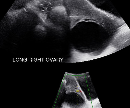
The radiologic evaluation of ultrasound showed an enlarged right ovary with a large follicular cyst and decreased blood flow in the pedicle. On Doppler ultrasound arterial blood flow may or may not be seen. Overall outcomes are good. The laboratory evaluation showed she was not pregnant, her white blood cell count was 11.5