This site uses cookies to improve your experience. To help us insure we adhere to various privacy regulations, please select your country/region of residence. If you do not select a country, we will assume you are from the United States. Select your Cookie Settings or view our Privacy Policy and Terms of Use.
Cookie Settings
Cookies and similar technologies are used on this website for proper function of the website, for tracking performance analytics and for marketing purposes. We and some of our third-party providers may use cookie data for various purposes. Please review the cookie settings below and choose your preference.
Used for the proper function of the website
Used for monitoring website traffic and interactions
Cookie Settings
Cookies and similar technologies are used on this website for proper function of the website, for tracking performance analytics and for marketing purposes. We and some of our third-party providers may use cookie data for various purposes. Please review the cookie settings below and choose your preference.
Strictly Necessary: Used for the proper function of the website
Performance/Analytics: Used for monitoring website traffic and interactions
Yes, we’re talking about your clavicular , proximal humeral, supracondylar, lateral condylar , scaphoid and metacarpal fractures. Today, we want to focus on a couple of our good friends, buckle and greenstick forearm fractures. Pediatric patients have unique bony anatomy and physiology compared to the skeletally mature.
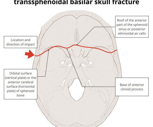
Recently, while reading an autopsy report, I ran across the term “hinge fracture of the skull.” A hinge fracture crosses the skull base transversely and involves the temporal and sphenoid bones. Here are diagrams of two common transsphenoidal fracture patterns, courtesy of radiopaedia.org. ” What?
Here are the factoids: Only 110 patients had a postop CT scan; 73 had scans within the first 24 hours, the other 37 were scanned later The rationale for early scan was to investigate retroperitoneal injury in half of patients, but frequently no indication was given (41%) The rationale for late scan was for workup of ileus in one-third or for evaluation (..)
We have previously discussed how the surrounding ligaments and tendon are often stronger than the weakest part of the child’s bone necessitating our vigilance when addressing the pediatric extremity complaint (ex, Ankle Pain , Elbow Injury , Supracondylar Fractures ). Of course, we cannot assume all anterior knee pain is benign.
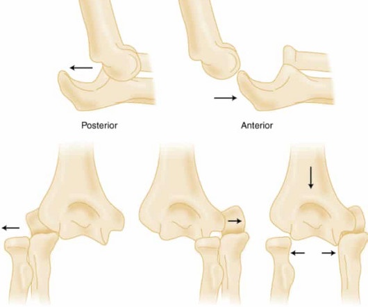
Elbow Dislocation Definition: Disarticulation of the proximal radius & ulna bones from the humerus Epidemiology: Incidence Second most common joint dislocation (after shoulder) in adults Most commonly dislocated joint in children Accounts for 10-25% of all injuries to the elbow ( Cohen 1998 ) Posterolateral is the most common type of dislocation (..)
Earlier this year, I wrote a series of posts on the two commonly used pelvic fracture interventions: preperitoneal packing (PPP) and angioembolization (AE). Patients with pelvic fractures are already at high risk for it. But what about venous thromboembolism risk?
Ultrasonography or radiography for suspected pediatric distal forearm fractures. Casey currently splits his time […] The post SGEM#415: Buckle Down for some Ultrasound to Diagnosis Distal Forearm Fractures first appeared on The Skeptics Guide to Emergency Medicine. Reference: Snelling et al. He is also a fully-fledged ultrasonographer.
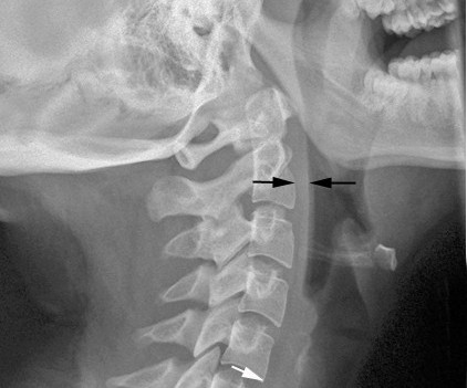
Patients with cervical fractures more commonly need a tracheostomy for ventilatory support and/or have a head injury , and these are well known culprits in dysphagia Normal soft tissue (<6mm at C2, <22mm at C6) A study in the Jan 2011 Journal of Trauma outlined the dysphagia problem seen with placement of a halo vest.
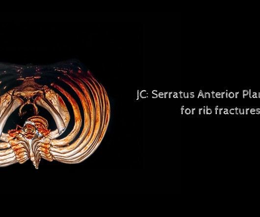
stemlyns #FOAMed The post JC: Serratus Anterior Plane Blocks for rib fractures in the ED. St.Emlyn's - Emergency Medicine #FOAMed JC: Review of an RCT of serratus anterior plane blocks in the emergency department for chest/rib injury. Is this now a standard of care? St Emlyn’s appeared first on St.Emlyn's.
Which clinical features best predict occult scaphoid fractures? He will soon be transitioning out of the US military after […] The post SGEM#420: I get knocked down, but I get up again – do I have a scaphoid fracture? Which clinical features best predict occult scaphoid fractures? Emerg Med J. Emerg Med J. Emerg Med J.
The foot X-Ray taken of the foot of the patient with the foot problem The Diagnosis She has a fracture at the base of the 5th metatarsal (Jones fracture). I like to think of these injuries as the scaphoid fracture of the foot, as this location of the metatarsal in particular has relatively poor blood supply. Am Fam Physician.
University of Maryland Department of Emergency Med
JULY 8, 2023
Pelvic fractures can be a major source of life threatening hemorrhage. Suspect fracture with significant force/mechanism. Signs are pelvic tendern. Click to view the rest
The foot X-Ray taken of the foot of the patient with the foot problem The Diagnosis She has a fracture at the base of the 5th metatarsal (Jones fracture). I like to think of these injuries as the scaphoid fracture of the foot, as this location of the metatarsal in particular has relatively poor blood supply. Am J Sports Med.
This places a significant responsibility on the EM physician to diagnose and treat fractures. Specifically, EM physicians should be able to recognize fractures that will likely require operative management and facilitate close follow up, such as a Maisonneuve fracture. 2) It is a specific type of a Weber C ankle fracture.
You may be shocked to […] The post JC: Evaluation of Lidocaine Patches for Elderly Patients with Rib Fractures: A Feasibility Study appeared first on St.Emlyn's.
Background Lidocaine patches, applied over rib fractures, may reduce pulmonary complications in older patients. We aimed to establish whether a definitive randomised controlled trial (RCT) evaluating lidocaine patches in older patients with rib fracture(s) was feasible.
You may be shocked to hear that an Emergency Medicine doctor such […] The post Lidocaine Patches for Elderly Patients with Rib Fractures: A Feasibility Study appeared first on St.Emlyn's. St.Emlyn's - Emergency Medicine #FOAMed Background I spend some of my time as a major trauma consultant on the major trauma ward (MTW).
Please consider a donation to ensure EM Cases continues to provide you high quality Free Open Access Medical Education here: [link] The post EM Quick Hits 60 Post-Tonsillectomy Hemorrhage, Post-CABG Infections, Bougie Tips, Pelvic Fracture Bleeds, Debriefing: Why, When & How appeared first on Emergency Medicine Cases.
Many vertebral fractures can be treated non-operatively. So I would like to concentrate on some papers that examined the use of back braces on patients who underwent pedicle screw fixation of their thoracic and/or lumbar spine fractures. Is postoperative bracing after pedicle screw fixation of spine fractures necessary?
She has pain on the radial aspect of her left wrist and anatomical snuffbox.However, the X-rays do not indicate an obvious scaphoid fracture. Clinical question: What are the predictive clinical features for occult scaphoid fractures in patients with normal initial radiographs in the ED?
Ultrasound‐Guided Serratus Anterior Plane Block (SAPB) Improves Pain Control in Patients With Rib Fractures. This topic is important because of the high incidence of rib fractures in trauma patients and their association with increased morbidity and mortality. Serratus Anterior Plane Blocks for Rib Fractures in the ED.
Problems can also arise when the tibia is fractured, leading to leakage into the soft tissues. But complications are possible. The most common is an insertion “miss”, where the fluid then infuses into the knee joint or soft tissues of the leg. Infection is extremely rare.
Efficacy and safety of ultrasound-guided erector spinae plane block compared to sham procedure in adult patients with rib fractures presenting to the emergency department: A randomized controlled trial. His imaging shows mildly displaced rib fractures of ribs four through seven. Reference: Ramesh S, Ayyan SM, Rath DP, Sadanandan DM.
Introduction Clinical Definition An open fracture is when the broken bone breaks through the skin or any other body cavity that is open to the outside, including those through the rectum or vagina. This is especially vital when addressing the issue of a fracture, adequate soft tissue coverage or blood vessel injury.
Danny McGurgan and Mike Cadogan Galeazzi fracture Galeazzi fracture (1934). Fracture of the distal third of the radius with associated Distal radio-ulna joint (DRUJ) disruption.
In this series, I will review the two major techniques for addressing troublesome bleeding from pelvic fractures. A multi-center trial published in 2015 showed an astounding 32% mortality rate for patients with shock from pelvic fracture. As I continue to preach, going anywhere but the OR is dangerous for the patient.
Whether you’re interested in orthopaedics or not, knowledge of basic fracture management can be useful in any ED. Examination of a fractured limb Most patients will be in a lot of pain. If the limb is pale and pulseless this needs urgent referral to vascular as well as orthopaedics and the fracture needs reducing immediately.
St.Emlyn's - Emergency Medicine #FOAMed Day 2 of the London Trauma Conference delivered impactful discussions on trauma care innovations, including advanced resuscitation strategies, rib fracture management, and prehospital interventions.
The trauma group at Stanford paired up with the Chang Gung Memorial Hospital in Taiwan to test the use of AI for interpreting images to identify a specific set of common pelvic fractures. The algorithms generated a “heat map” that showed the areas that were suspicious for fracture. All were handily identified by the AI.
University of Maryland Department of Emergency Med
JUNE 24, 2023
Use of intravenous lidocaine has been proposed as an adjunct/replacement for opioids in trauma patients with rib fractures. These small st. Click to view the rest
[Pereira 2017] If pathologic fractures occur, may be misdiagnosed as non-accidental trauma. [Li The initiation of antibiotic therapy in some infants can lead to a Jarisch–Herxheimer reaction (fevers, chills, hypotension, and possibly fetal death) due to the intense inflammatory reaction of the body to dying spirochetes.
In this intro video, we demonstrate the use of ultrasound imaging to identify fractures. Ultrasound-guided fracture identification is a minimally invasive technique that allows for quick and accurate diagnosis of fractures, without the need for radiation or contrast agents. Lrg Joint Tap Basic Knee Exam Shoulder Exam
Specific ones that come to mind are shock, long bone or spine fractures, and TBI. Similarly, was there a relationship between the number of units of whole blood and possible “protection” from VTE? Did you examine other physiologic or anatomic variables and their relationship with VTE?
Serratus Anterior Plane Blocks for Early Rib Fracture Pain Management: The SABRE Randomized Clinical Trial. He has three left-sided rib fractures on imaging without underlying complications. Background: Rib fractures are a common injury, particularly for those over 65. We have covered rib fractures twice on the SGEM.
Let's review some high yield aspects of rib fractures and the most simplified approach you will ever hear. Rib fractures: T-giving football. Let's review some high yield aspects of rib fractures and the most simplified approach you will ever hear. Rib fractures: T-giving football. Cite this podcast as: Briggs, Blake.
Let's review some high yield aspects of rib fractures and the most simplified approach you will ever hear. Rib fractures: T-giving football. Let's review some high yield aspects of rib fractures and the most simplified approach you will ever hear. Rib fractures: T-giving football. Cite this podcast as: Briggs, Blake.
They have thinner bones of the skull which makes them more prone to skull fractures. of these had a CT performed 1 (0.2%) had a clinically important TBI (ciTBI) 10 (5.1%) had any TBI on CT 9 (4.6%) had skull fractures Remember to always watch out for NAT ! PECARN is highly sensitive for identifying those with clinically important TBI.
Hamate fractures are uncommon, but early and accurate diagnosis is critical to prevent negative outcomes. Hamate fractures may present initially to emergency departments, and diagnosis can be challenging.
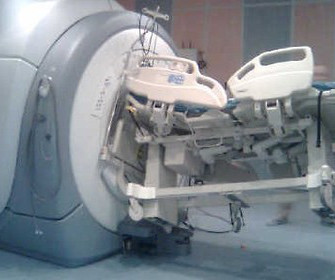
This is typically a problem that arises in head-injured patients with extremity or pelvic fixators for concomitant fractures. I’ve covered the problem of performing MRI on patients with external fixators. MRI is an indispensable tool for the evaluation of head, spine, and soft tissue trauma.
In this episode of the ETM Course Podcast we talk to Dr Chris Partyka, Emergency Physician and Prehospital and Retrieval Specialist from Sydney and lead author for the recently published SABRE trial which compared Serratus Anterior Plane Blocks to a standard analgesia package for patients with rib fractures.
What is a Hairline Fracture? A common cause of sports injuries, hairline fracture is a type of fracture that begins as a minor inflammation on the surface of a bone and manifests as a tiny thin hair-like crack on the bone. It is also known as a stress fracture or a fissure fracture.
Loss of cervical lordosis Concerning mechanism Remember: CT will help identify bony injury, however MRI is the better test to assess for ligamentous injury as well as spinal cord injury Below is an example of a Hangman’s Fracture (See Cervical Spine Fractures ). Moral of the Morsel You eyes may deceive you!
We organize all of the trending information in your field so you don't have to. Join 5,000+ users and stay up to date on the latest articles your peers are reading.
You know about us, now we want to get to know you!
Let's personalize your content
Let's get even more personalized
We recognize your account from another site in our network, please click 'Send Email' below to continue with verifying your account and setting a password.


Let's personalize your content