This site uses cookies to improve your experience. To help us insure we adhere to various privacy regulations, please select your country/region of residence. If you do not select a country, we will assume you are from the United States. Select your Cookie Settings or view our Privacy Policy and Terms of Use.
Cookie Settings
Cookies and similar technologies are used on this website for proper function of the website, for tracking performance analytics and for marketing purposes. We and some of our third-party providers may use cookie data for various purposes. Please review the cookie settings below and choose your preference.
Used for the proper function of the website
Used for monitoring website traffic and interactions
Cookie Settings
Cookies and similar technologies are used on this website for proper function of the website, for tracking performance analytics and for marketing purposes. We and some of our third-party providers may use cookie data for various purposes. Please review the cookie settings below and choose your preference.
Strictly Necessary: Used for the proper function of the website
Performance/Analytics: Used for monitoring website traffic and interactions
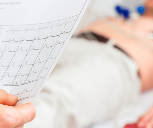
Electrocardiograms (EKGs) are at the top of modern healthcare as indispensable diagnostic tools for identifying arrhythmias, heart attacks, and other cardiac disorders. In this blog, we delve into the power of EKG training, the essential skills healthcare professionals need, and strategies to excel in using this life-saving technology.
I have often written about how an ECG interpreted as "normal" by a conventional algorithm may well be manifesting OMI, or even long QT or hyperkalemia. Shifa Karim and Gabe Keller helped with a project to assess all these ECGs with the Queen of Hearts. The ECG told the story. I wanted to show some of the cases here.
This ECG was recorded: What do you think? EKG showed sinus rhythm at 100 BPM with a normal axis and diffuse ST depressions. Another ECG was recorded: What do you think? Not knowing anything about the patient, I saw this ECG on our system just as I was leaving the dept. There is widespread ST depression.
I want all to know that, with the right mind preparation, and the use of the early repol/LAD occlusion formula, extremely subtle coronary occlusion can be detected prospectively, with no other information than the ECG. His ECG was repeated at this point: This shows a well developed anterior STEMI. His first troponin was normal.
We organize all of the trending information in your field so you don't have to. Join 5,000+ users and stay up to date on the latest articles your peers are reading.
You know about us, now we want to get to know you!
Let's personalize your content
Let's get even more personalized
We recognize your account from another site in our network, please click 'Send Email' below to continue with verifying your account and setting a password.


Let's personalize your content