

Chest Pain in Children: ReBaked Morsel
Pediatric EM Morsels
MAY 10, 2024
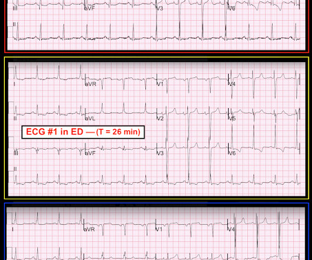
EKG Reasonable screen for cardiac etiology [ Kane, 2010 ]: Chest Pain with Exertion? Ultrasound diagnosis of occult pneumothorax. The role of point-of-care ultrasound in the diagnosis of pericardial effusion: a single academic center retrospective study. Ultrasound J. Abnormal exam (ex, murmurs, hepatomegaly)?


Let's personalize your content