Remove EKG/ECG Remove Stroke Remove Wellness
article thumbnail

ECG Blog #435 — Did Cath Show Acute Ischemia?

Ken Grauer, MD

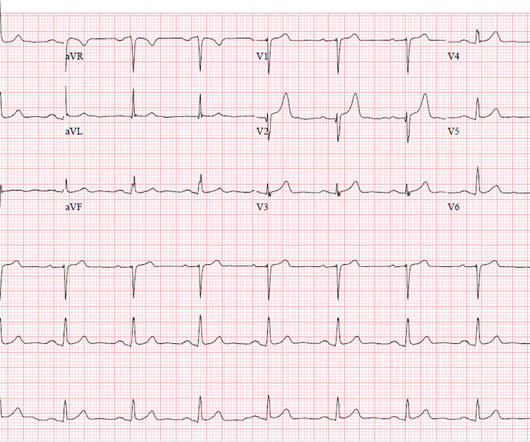
The ECG in Figure-1 — was obtained from a middle-aged woman with positional tachycardia and diaphoresis with change of position from suprine to sitting. Although CP ( C hest P ain ) was not a prominent symptom — ACS ( A cute C oronary S yndrome ) was suspected from the chest lead T wave inversion seen on this ECG. WHY — or Why Not?

EKG/ECG 433
article thumbnail

The Study of the Week Is a Beautiful Example of Science Done Well

Sensible Medicine

Neurologist Hooman Kamel from the Weil Cornell Medical Center in NY had an idea about atrial fibrillation and stroke. Stroke came when these clots moved northward to the brain. One of the big issues with this theory was that studies had failed to show a strong relationship in time between the stroke and the irregular rhythm. (For

Wellness 123
Insiders

Sign Up for our Newsletter

This site is protected by reCAPTCHA and the Google Privacy Policy and Terms of Service apply.

article thumbnail

An elderly male with acute altered mental status and huge ST Elevation

Dr. Smith's ECG Blog

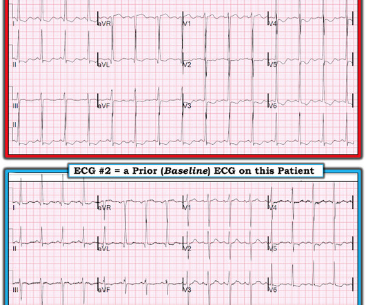
CTA head and neck were obtained and showed no evidence of intracranial hemorrhage, large vessel occlusion stroke (what a helpful and apt name for an acute arterial occlusion paradigm, by the way.), EKG on arrival to the ED is shown below: What do you think? 2) There was no terminal QRS distortion on these ECGs. Any changes?

EKG/ECG 103
article thumbnail

A 90-something with acute stroke. She has no chest symptoms. What is the diagnosis?

Dr. Smith's ECG Blog

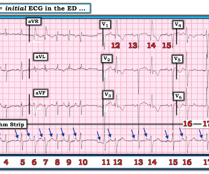
A 90-something year old woman presented with an acute mild stroke. She had a routine ECG as part of her workup: What do you think? Peak trop 62 ng/L (would be very low for acute OMI) Next AM ECG: Still with very ischemic looking T-waves. Always look at old ECGs, even if you think the diagnosis is obvious.

Stroke 70
article thumbnail

A man in his 60s with syncope and ST depression. What does the ECG mean?

Dr. Smith's ECG Blog

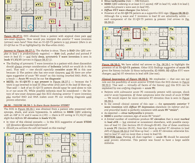
Here is his triage ECG: What do you think? What is the most likely cause of the patient’s ECG findings, and what would be your first step in management? What does the ECG show? What does the ECG show? Figure-1: The initial ECG in this case — and a rapid method for estimating the QTc ( See text ). Is it STEMI?

EKG/ECG 117
article thumbnail

STREAM-2: Half-Dose Tenecteplase vs Primary PCI in Older Patients with STEMI?

RebelEM

ECG Results: Repeat ECG 90min after tenecteplase indicated 70.3% ECG Results: Repeat ECG 90min after tenecteplase indicated 70.3% ECG Results: Repeat ECG 90min after tenecteplase indicated 70.3% The upper range of this time was 120 minutes from diagnostic ECG in the majority of patients.

EKG/ECG 138
article thumbnail

AF Duration? Another Big Story from 2023

Stop and Think

For an older patient with stroke risk factors and 3 hours of AF on a cardiac device, pre-2023 thinking would have us leaning toward anticoagulation. Edoxaban reduced the primary endpoint of stroke, systemic embolism, and cardiovascular death by 19% (HR, 0.81; 95% CI, 0.60-1.08; The doctor would do an ECG and that would diagnose AF.

Stroke 90