
ECG Blog #443 — A 40s Man with CP and Dyspnea
Ken Grauer, MD
AUGUST 16, 2024
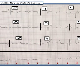
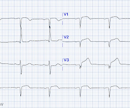
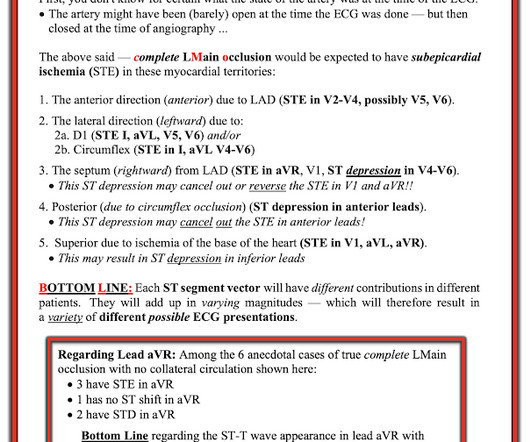
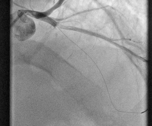
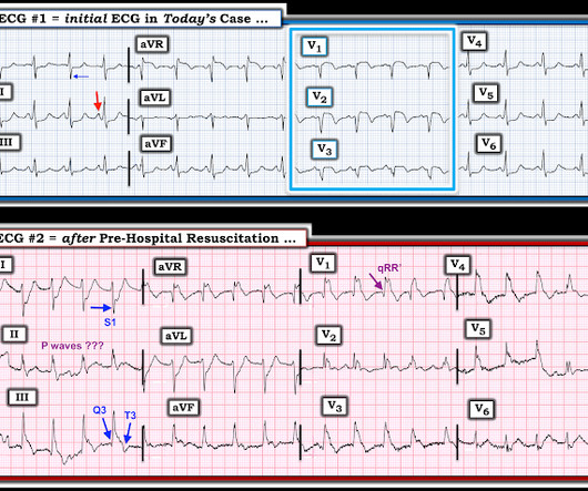
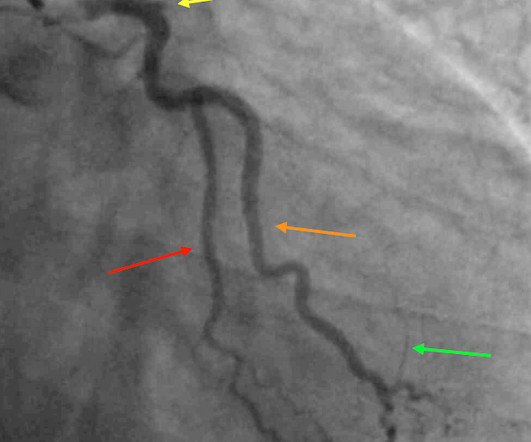
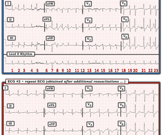
The ECG in Figure-1 was obtained from a man in his 40s — who presented to the ED ( E mergency D epartment ) because of CP ( C hest P ain ) and shortness of breath. QUESTIONS: In view of the above history — How would YOU interpret the ECG in Figure-1 ? Based on the history and the patient's initial ECG — the cath lab was activated.


Let's personalize your content