Remove EKG/ECG Remove Shock Remove Ultrasounds
article thumbnail

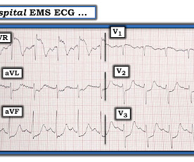
Another deadly triage ECG missed, and the waiting patient leaves before being seen. What is this nearly pathognomonic ECG?

Dr. Smith's ECG Blog

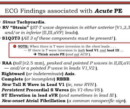
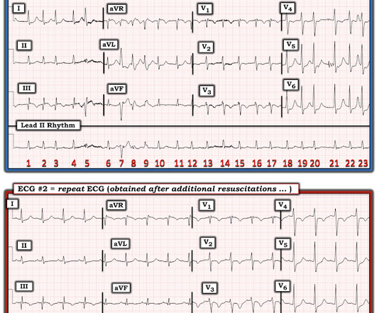
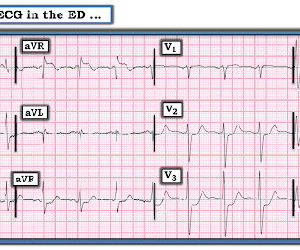
Pain improved to 1/10 after EMS administers 324 mg aspirin and the following EKG is obtained at triage. If this EKG were handed to you to screen from triage without any clinical information, what would you think? Do you appreciate any dynamic changes compared to the patient’s prior EKG? What do you think? In fact, Kosuge et al.

EKG/ECG 137
article thumbnail

Chest pain and shock: Is there a right ventricular OMI on this ECG? And should he undergo trancutaneous pacing?

Dr. Smith's ECG Blog

A 50-something man presented in shock with severe chest pain. His prehospital ECG was diagnostic of inferior posterior OMI. The patient was in clinical shock with a lactate of 8. Here is his ED ECG: There is bradycardia with a junctional escape. RVMI explains part of the shock. What is the atrial activity?

Shock 79
Insiders

Sign Up for our Newsletter

This site is protected by reCAPTCHA and the Google Privacy Policy and Terms of Service apply.

Trending Sources

article thumbnail

A man in his 50s with shortness of breath

Dr. Smith's ECG Blog

Here is his ECG: Original image, suboptimal quality Quality improved with PM Cardio digitization The ECG is highly suggestive of acute right heart strain, with sinus tachycardia, S1Q3T3, and T wave inversions in anterior and inferior with morphology consistent with acute right heart strain. Moreover, there is tachycardia.

EKG/ECG 71
article thumbnail

A 50-something with Regular Wide Complex Tachycardia: What to do if electrical cardioversion does not work?

Dr. Smith's ECG Blog

Initial ED ECG: What do you think? Then we must consider clinical data other than the ECG, for a pretest probability : Of all wide complex tachydysrhythmias, the majority are VT. Shocked x 2 without effect. Pads were placed with ultrasound guidance, so they were in the correct position. Why did I say that? What to do now?

EKG/ECG 136
article thumbnail

Noisy, low amplitude ECG in a patient with chest pain

Dr. Smith's ECG Blog

Colin is an emergency medicine resident beginning his critical care fellowship in the summer with a strong interest in the role of ECG in critical care and OMI. An ECG was obtained shortly after arrival: ECG 1 What do you think? The ECG has a lot of artifact, and the amplitude is very small, making interpretation challenging.

EKG/ECG 66
article thumbnail

Extreme shock and cardiac arrest in COVID patient

Dr. Smith's ECG Blog

He had the following EKG recorded: Low voltage, suggests effusion. A bedside cardiac ultrasound was normal, with no effusion. He underwent CPR, and regained a pulse after epinephrine, with an organized narrow complex rhythm at 140, but still with severe shock. Assessment was severe sudden cardiogenic shock. What is it?

Shock 52
article thumbnail

Severe shock, obtunded, and a diagnostic prehospital ECG. Also: How did this happen?

Dr. Smith's ECG Blog

She was in shock with thready pulses. A prehospital ECG was recorded: Limb leads: Precordial Leads What is the therapy? The rate is not fast enough to be causing shock, so if it is VT, the priority is still to treat hyperK and secondarily to cardiovert. They thought it was VT, but did not shock. The K returned at 7.4

Shock 52