Remove EKG/ECG Remove Sepsis Remove Wellness
article thumbnail

ECG Blog #451 — Premature Closure.

Ken Grauer, MD

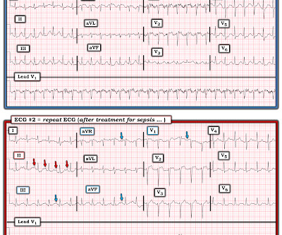
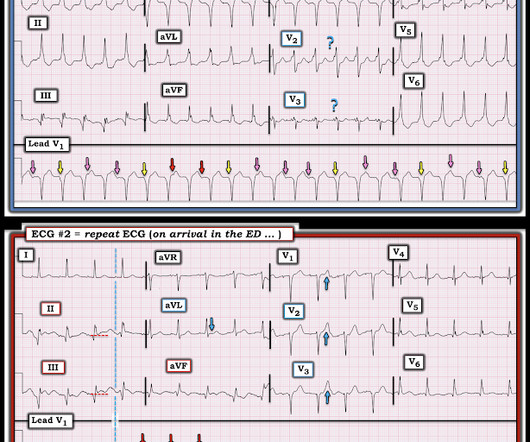
I was sent the ECG shown in Figure-1 — told only that the patient was a middle-aged man with septicemia. Figure-1: The initial ECG in today's case. With practice — it should literally take no more than seconds to assess these 5 Parameters ( See ECG Blog #185 — for more on the Ps,Qs,3R Approach to rhythm interpretation ).

EKG/ECG 418
article thumbnail

Torsade in a patient with left bundle branch block: is there a long QT? (And: Left Bundle Pacing).

Dr. Smith's ECG Blog

She was managed for sepsis with antibiotics including azithromycin, had hypotension with arterial and central lines placed and pressors. She had an ECG recorded: This is left bundle branch block (LBBB), with appropriate proportional discordance. Bedside cardiac ultrasound showed moderately decreased LV function. J Am Coll Cardiol.

EKG/ECG 115
Insiders

Sign Up for our Newsletter

This site is protected by reCAPTCHA and the Google Privacy Policy and Terms of Service apply.

Trending Sources

article thumbnail

ECG Blog #366 — Diltiazem didn't work.

Ken Grauer, MD

The ECG and long lead II rhythm strip in Figure-1 — was obtained from a COVID positive patient with persistent tachycardia not responding to Diltiazem. Figure-1: The initial ECG — obtained from a patient with persistent tachycardia. ( To improve visualization — I've digitized the original ECG using PMcardio ).

EKG/ECG 195
article thumbnail

Putting Clinical Gestalt to Work in the Emergency Department

ACEP Now

For example, experienced emergency physicians have great clinical gestalt and accuracy to predict sepsis in critically ill patients at just 15 minutes from patient arrival—more so than scoring tools like the qSOFA, MEWs, and even machine-learning trained artificial intelligence models.

article thumbnail

Grand Rounds Recap 3.6.24

Taming the SRU

Soon you’ll be able to view our whole pathway on the QI/KT page, but for now, we hope everyone remembers these points: 1) Get labs and an EKG on everyone presenting with hypoglycemia. ECG is a must for EVERY syncope patient. It is not specific enough in defining specific findings on the ECG that should be considered high risk.

EKG/ECG 98
article thumbnail

Tachycardia must make you doubt an ACS or STEMI diagnosis; put it all in clinical context

Dr. Smith's ECG Blog

He had this ECG recorded. He was rushed by residents into our critical care room with a diagnosis of STEMI, and they handed me this ECG: There is sinus tachycardia with ST elevation in II, III, and aVF, as well as V4-V6. But, remember, we do not evaluate and treat ECGs, we evaluate and treat patients. Are the lungs clear?

EKG/ECG 52
article thumbnail

What clinical scenario fits best?

Dr. Smith's ECG Blog

B elow are 4 ECGs from the same patient. Accompanying the ECGs is some clinical information. Look at the ECGs and consider the timeline and other information. Which one do you think fits better with the ECGs and their timeline? As part of her work up in the ED an ECG was recorded. ECG#2 - day 4.

EKG/ECG 110