Remove EKG/ECG Remove Sepsis Remove Shock
article thumbnail

ECG Blog #366 — Diltiazem didn't work.

Ken Grauer, MD

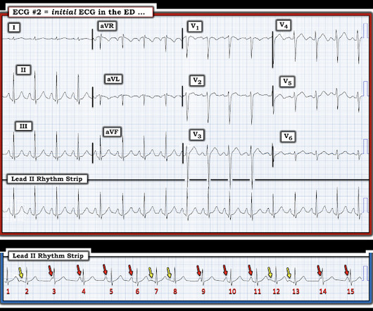
The ECG and long lead II rhythm strip in Figure-1 — was obtained from a COVID positive patient with persistent tachycardia not responding to Diltiazem. Figure-1: The initial ECG — obtained from a patient with persistent tachycardia. ( To improve visualization — I've digitized the original ECG using PMcardio ).

EKG/ECG 195
article thumbnail

See what happens when a left main thrombus evolves from subtotal occlusion to total occlusion.

Dr. Smith's ECG Blog

The ECG below was recorded about 20 minutes after he regained consciousness. ECG #1 The above ECG shows sinus rhythm at about 60 bpm. Figure A It now becomes apparent that there is ST segment depression in almost every lead of the ECG (V1-V6, I, II, aVL and aVF). sepsis, anemia, hypoxemia, severe hypotension etc.,

EKG/ECG 125
Insiders

Sign Up for our Newsletter

This site is protected by reCAPTCHA and the Google Privacy Policy and Terms of Service apply.

Trending Sources

article thumbnail

Episode 7: Sepsis

PHEM Cast

[link] We hope you enjoyed our sepsis podcast. St Emlyns Induction podcast on Sepsis. A great summary of what to do when a patient with suspected sepsis first arrives in the ED. A great summary of what to do when a patient with suspected sepsis first arrives in the ED. March 2016.

Sepsis 52
article thumbnail

Grand Rounds Recap 3.6.24

Taming the SRU

Soon you’ll be able to view our whole pathway on the QI/KT page, but for now, we hope everyone remembers these points: 1) Get labs and an EKG on everyone presenting with hypoglycemia. ECG is a must for EVERY syncope patient. It is not specific enough in defining specific findings on the ECG that should be considered high risk.

EKG/ECG 95
article thumbnail

Tachycardia must make you doubt an ACS or STEMI diagnosis; put it all in clinical context

Dr. Smith's ECG Blog

He had this ECG recorded. He was rushed by residents into our critical care room with a diagnosis of STEMI, and they handed me this ECG: There is sinus tachycardia with ST elevation in II, III, and aVF, as well as V4-V6. But, remember, we do not evaluate and treat ECGs, we evaluate and treat patients. Are the lungs clear?

EKG/ECG 52
article thumbnail

emDOCs Podcast – Episode 93: BRASH Syndrome

EMDocs

Episode 93: BRASH syndrome Background: Brash syndrome has 5 components: bradycardia, renal failure, AV nodal blocker, shock, hyperkalemia. An inciting event will typically push them over the edge into BRASH syndrome: Dehydration Hypotension from sepsis or another condition GI illness Dosage increase of a chronic medication (e.g.,

Shock 87
article thumbnail

What are treatment options for this rhythm, when all else fails?

Dr. Smith's ECG Blog

The below ECG was recorded. The ECG shows obvious STEMI(+) OMI due to probable proximal LAD occlusion. This ECG does not have the typical ST-vector of an LAD occlusion. See below for Ken Grauer Comment on the initial ECG: == On arrival, another ECG was recorded: There appears to have been quite a bit of spontaneous reperfusion!

EKG/ECG 119