Remove EKG/ECG Remove Resuscitation Remove Ultrasounds
article thumbnail

A 50-something with chest pain.

Dr. Smith's ECG Blog

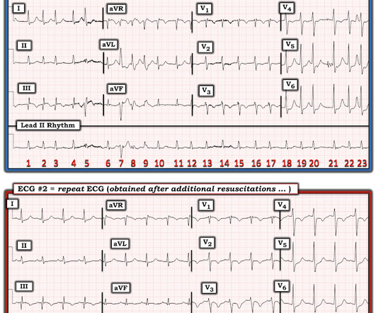
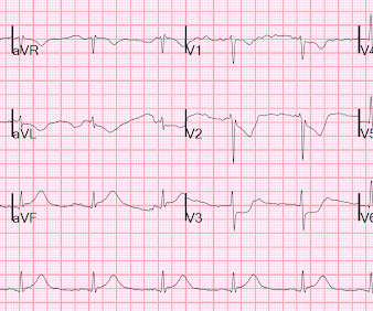
This ECG was recorded in triage. The computer interpretation is: “Sinus Brady with moderate intraventricular conduction delay, nonspecific t wave abnormality, abnormal EKG” What do you think? Case Continued The ECG findings were not recognized. Resuscitative attempts were initiated quickly. LCX with moderate disease.

EKG/ECG 128
article thumbnail

Which patient needs a CT scan?

Dr. Smith's ECG Blog

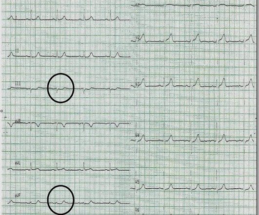
She had this ECG recorded: Obvious massive anterior STEMI She was quickly brought to the critical care area and the cath lab was activated. No ECG was recorded after pain resolution. Here is the ECG at 25 minutes: Terrible LAD STEMI (+) OMI So a CT scan was done which of course showed a normal aorta. She was a walk-in at triage.

Insiders

Sign Up for our Newsletter

This site is protected by reCAPTCHA and the Google Privacy Policy and Terms of Service apply.

Trending Sources

article thumbnail

A man in his 30s with cardiac arrest and STE on the post-ROSC ECG

Dr. Smith's ECG Blog

Initial ROSC was obtained, during which this ECG was obtained: What do you think? I sent it to 2 of my ECG nerd colleagues with no clinical information whatsoever, who instantly said: "Looks like afib with subendocardial ischemia and right heart strain pattern." "I CPR was initiated immediately. The rhythm is atrial fibrillation.

EKG/ECG 52
article thumbnail

emDOCs Podcast – Episode 98: Post ROSC Mental Model

EMDocs

Check the pulse RSI= Resuscitation Sequence Intubation Hypoxia, Hypotension, and Acidosis are the reason patients code during/post intubation These patients are super high risk for all 4 Optimize first pass success – Induction agent + paralytic Unconscious patients will still have muscle tone Induction Ketamine or Etomidate at half doses (i.e.,

article thumbnail

First10EM Journal Club: March 2024

Broome Docs

If you want to see Justin in the video format and his new Youtube series then click here: [link] If you would like to check out my current ultrasound training project then click here: WACHS Ultrasound Otherwise – enjoy the show and see you in a few weeks. Myth 3: The ECG changes from hyperkalemia are predictable and reliable.

PPE 107
article thumbnail

Tachycardia must make you doubt an ACS or STEMI diagnosis; put it all in clinical context

Dr. Smith's ECG Blog

He had this ECG recorded. He was rushed by residents into our critical care room with a diagnosis of STEMI, and they handed me this ECG: There is sinus tachycardia with ST elevation in II, III, and aVF, as well as V4-V6. But, remember, we do not evaluate and treat ECGs, we evaluate and treat patients. The K returned at 6.9

EKG/ECG 52
article thumbnail

Cardiac arrest, LBBB with STEMI on the ECG, but no Acute Coronary Syndrome!

Dr. Smith's ECG Blog

He was resuscitated after fairly prolonged down time, but regained consciousness, though he was confused. Here is the prehospital ECG at 1935: Sinus rhythm with left bundle branch block (LBBB). In Sgarbossa's study, just 1 mm concordant STE in just 1 lead was 92% specific for MI and earned the ECG 5 points.

EKG/ECG 52