article thumbnail

ECG Blog #394 — Is QRS Morphology Disguised?

Ken Grauer, MD

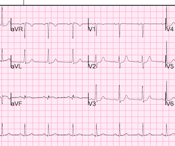
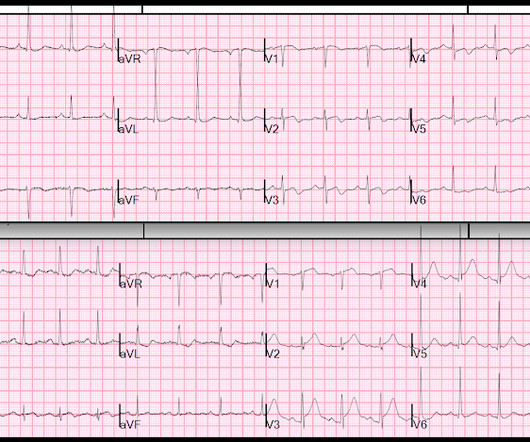
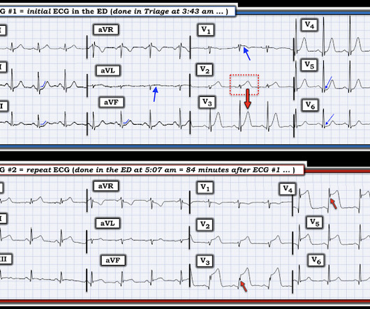
The ECG in Figure-1 — was obtained from an older man with a history of prior infarction and coronary bypass surgery. Figure-1: The ECG in today's case. ( To improve visualization — I've digitized the original ECG using PMcardio ). There is no ECG indication of chamber enlargement — ST-T wave changes do not look acute.

EKG/ECG 292
article thumbnail

What makes a T-wave Hyperacute? And: 30 Examples of Hyperacute T-waves, 10 in each of 3 myocardial territories.

Dr. Smith's ECG Blog

The way to get good at it is to see a lot of them, and also see a lot of fake HATWs (mimics) Here is a difficult pair of ECGs that demonstrate a difference: One ECG is normal variant STE. The more abnormal leads and lead areas you can identify in a given ECG — the more solid the evidence of acute OMI becomes. Which is which?

EKG/ECG 133
Insiders

Sign Up for our Newsletter

This site is protected by reCAPTCHA and the Google Privacy Policy and Terms of Service apply.

article thumbnail

ToxCard: Second Generation Antipsychotic Overdose

EMDocs

Recommend obtaining multiple EKGs to aid in the diagnosis of cardiotoxic effects such as dysrhythmias or interval widening, even if not apparent immediately after the overdose. 1,2 Cardiovascular: Obtain an initial EKG to determine the patient’s baseline and repeat. 1,2 Consider lidocaine if sodium bicarbonate fails. 1 Class IA (e.g.,

Poisoning 108
article thumbnail

A 40-something male with resolving chest pain and a "Normal ECG" by computer and cardiology overread

Dr. Smith's ECG Blog

Here is his initial ED ECG: What do you think? I read this ECG without any history as reperfusing inferior and posterior OMI due to the Q-wave in lead III with minimal STE and reciprocal ST depression in V2-V4 (which should never be there). We send each other EKG by the dozens every day. Most are OMI look alikes (mimics).

EKG/ECG 95
article thumbnail

Lecture by Smith: Selected Cases of Occlusion MI (OMI), or not, on the ECG

Dr. Smith's ECG Blog

are compellingly answered by the several minute intro of his credentials and research in the field of acute ECG interpretation for suspected infarction — that I suspect are without parallel by anyone anywhere in the field of cardiology. So — FREEZE the picture of each ECG on the screen — FORCE yourself to commit to an answer.

EKG/ECG 98
article thumbnail

Cardiac Rhythms/ECG Module

Don't Forget the Bubbles

Add into this that the majority of children will be in normal sinus rhythm (NSR) by the time of assessment so to truly identify those who have something wrong we have to be confident in identifying arrhythmias where they are present and critical when analysing an ECG in NSR. All were examined and 98% had an ECG.

EKG/ECG 98
article thumbnail

An elderly male with shortness of breath

Dr. Smith's ECG Blog

ECG 1 at time zero EARLY REPOLARIZATION ABNORMAL ECG ED final official overread: "early repol vs hyperacute T, minimal changes from previous (previous shown below)" What do YOU think? Thus, the LAD has reperfused ("recanalized") spontaneously Queen on ECG 2: Not OMI, Low confidence She is also worried about V2.

EKG/ECG 117