Remove EKG/ECG Remove Radiology Remove Shock
article thumbnail

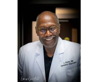
Dr. Elsburgh Clarke Was Among First to Specialize in Emergency Medicine

ACEP Now

A closer look, though, also shows the technology of the daya bulky, two-way radio for communicating with EMS, metal gurneys, glass saline bottles, and portable ECG monitors the size of a small shopping cart. Notice the use of the medical anti-shock trousers and the ECG machine. Click to enlarge.) I like the excitement.

article thumbnail

Medical Malpractice Insights: Failed communication of incidental mediastinal mass

EMDocs

The EKG and troponin are normal. He and his physician are shocked at the report and the lack of follow-up. A mediastinal mass – or any “OMG” imaging result warrants discussion with radiology and sharing the information with the patient. Exam is unremarkable. D-dimer is elevated at 1.6. Verbal communication is not enough.

Insiders

Sign Up for our Newsletter

This site is protected by reCAPTCHA and the Google Privacy Policy and Terms of Service apply.

article thumbnail

A crashing patient with an abnormal ECG that you must recognize

Dr. Smith's ECG Blog

Written by Pendell Meyers, with edits from Steve Smith Let's consider this nearly pathognomonic ECG without the clinical context (because sometimes the clinical context will not be as easy as in this case). This ECG is diagnostic of hemodynamically significant acute right heart strain. What is the answer? Why is it not Wellens???

EKG/ECG 40
article thumbnail

emDOCs Revamp: Esophageal Perforation

EMDocs

ECG – May show tachycardia or rate related ST depressions Laboratory evaluation: CBC w/ differential – may reveal leukocytosis with left shift CMP, Lipase – can reveal alternative intra-abdominal diagnoses as well as show findings of end-organ hypoperfusion (elevated serum creatinine, transaminitis, etc.)

article thumbnail

A woman in her 70s with chest pain

Dr. Smith's ECG Blog

Here is her ECG on arrival to the ED: What is your differential? The ECG is quickly reviewed and shows sinus rhythm with normal QRS complexes. Her prior ECG on file is shown below: What are your next steps? Learning Points: The ECG is always just one piece of the clinical puzzle. What are your next steps?

article thumbnail

Episode 29 - Assessing Abdominal Pain in Adults: A Rational, Cost-Effective, and Evidence-Based Strategy

EB Medicine

CT is good but you really should learn ultrasound, and lastly, sick patients need prompt consultation and resuscitation, not rapid trips to radiology. Nachi: Moving on to blood pressure: frank hypotension should make you immediately think of a ruptured AAA or septic shock 2/2 an intra abd infection. In one study, a SI > 0.7

article thumbnail

MI in Children

Pediatric Emergency Playbook

Electrocardiography (ECG) should be performed on any patient with significant blunt chest injury. A negative ECG is highly consistent with no significant blunt myocardial injury. Any patient with a new abnormality on ECG (dysrhythmia, heart block, or signs of ischemia) should be admitted for continuous ECG monitoring.