Remove EKG/ECG Remove Outcomes Remove Ultrasounds
article thumbnail

Chest pain, resolved. Does it need emergent cath lab activation (some controversy here)? And much much more.

Dr. Smith's ECG Blog

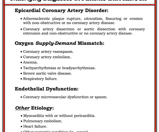
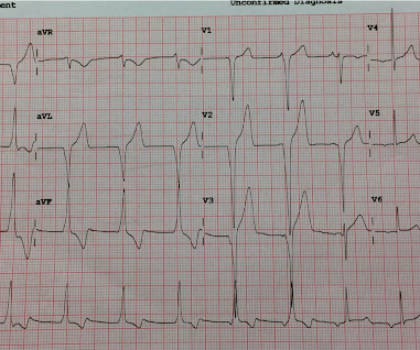
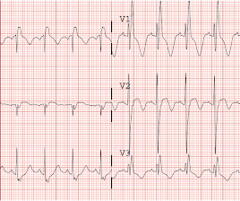
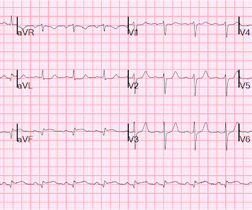
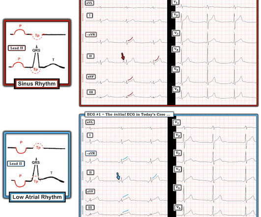
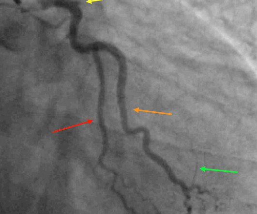
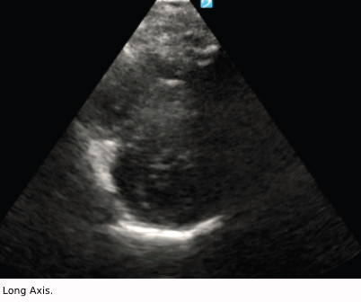
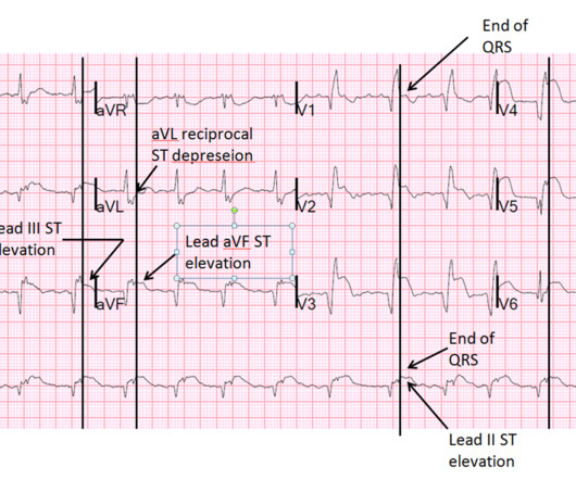
EKG from triage: Here is his previous ECG: Normal ST Elevation Resident's interpretation: Reperfusion pattern/Wellens' with biphasic T waves in V2 and V3, and in comparison to an EKG in 2020 this is new. Bedside ultrasound with no apparent wall motion abnormalities, no pericardial effusion, no right heart strain.

EKG/ECG 117
article thumbnail

Four patients with chest pain and ‘normal’ ECG: can you trust the computer interpretation?

Dr. Smith's ECG Blog

All initial ECGs were labeled ‘normal’ or ‘otherwise normal’ by the computer interpretation, and below are the ECGs with the final cardiology interpretation. 1-3] But these studies were very short duration and used cardiology interpretation of ECGs or emergent angiography rather than patient outcomes.

EKG/ECG 119
Insiders

Sign Up for our Newsletter

This site is protected by reCAPTCHA and the Google Privacy Policy and Terms of Service apply.

Trending Sources

article thumbnail

Putting Clinical Gestalt to Work in the Emergency Department

ACEP Now

Does that normal troponin and ECG obviate the need for cardiology consultation for my patient with a concerning story for acute coronary syndrome? 4 However, emergency physicians rely on gestalt to predict outcomes well. The “bundling” heuristic—if I send a troponin, then an ECG is needed. Urology took him for orchiopexy.

article thumbnail

Chest pain and shock: Is there a right ventricular OMI on this ECG? And should he undergo trancutaneous pacing?

Dr. Smith's ECG Blog

His prehospital ECG was diagnostic of inferior posterior OMI. Here is his ED ECG: There is bradycardia with a junctional escape. We recorded an ECG in which V1-V3 were put in the position of V4R-V6R, and V4-6 were placed in V7-9 to (academically) confirm posterior OMI. The patient was in clinical shock with a lactate of 8.

Shock 97
article thumbnail

Ventricular Fibrillation, ICD, LBBB, QRS of 210 ms, Positive Smith Modified Sgarbossa Criteria, and Pacemaker-Mediated Tachycardia

Dr. Smith's ECG Blog

Bedside ED ultrasound showed exceedingly poor global LV function, and no B lines. Here is the initial ED ECG. Other thought this was due to hyperkalemia, but the ECG does not have the appearance of hyperkalemia but does have the appearance of severe cardiomyopathy -- LBBB with very wide QRS) 3. What do you think?

EKG/ECG 132
article thumbnail

Grand Rounds Recap 1.3.24

Taming the SRU

R4 Case Follow-up: SCAD WITH dr. Martella Spontaneous Coronary Artery Dissection (SCAD) is a diagnosis confirmed via imaging: Coronary Angiography, Optical Coherence Tomography, Intravascular Ultrasound Therefore, treatment in the ED is the same as atherosclerotic ACS: ASA, heparin gtt and possible statin.

EKG/ECG 93
article thumbnail

Friday Reflection 41: Sometimes I Just Need to Complain

Sensible Medicine

There were no personal crises or untoward patient outcomes. The plan was that she would get a two-week, continuous EKG monitor placed and have a sleep study at this hospital. Last week was one of them. She follows up at a “highly ranked” west coast academic medical center, near where she lives half the year.

Stroke 106