

What makes a T-wave Hyperacute? And: 30 Examples of Hyperacute T-waves, 10 in each of 3 myocardial territories.
Dr. Smith's ECG Blog
NOVEMBER 27, 2024
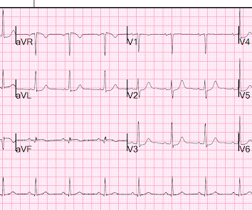
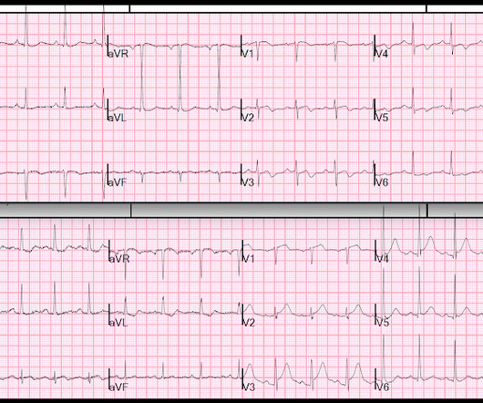
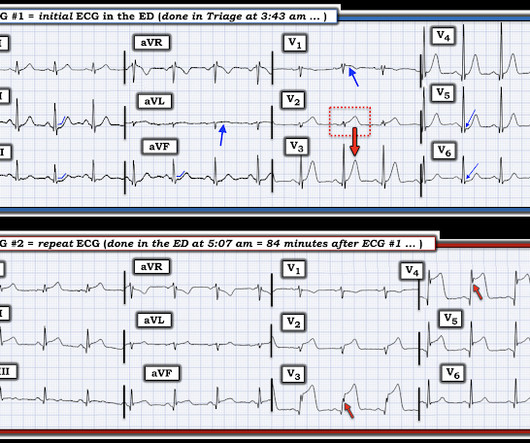
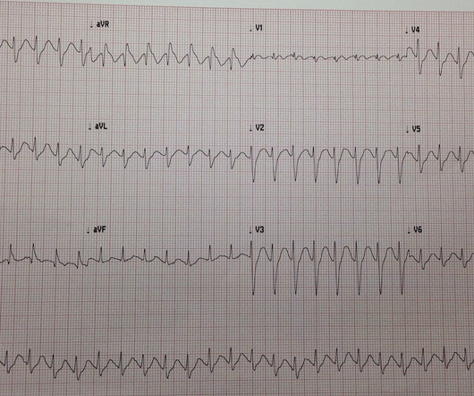
The way to get good at it is to see a lot of them, and also see a lot of fake HATWs (mimics) Here is a difficult pair of ECGs that demonstrate a difference: One ECG is normal variant STE. The more abnormal leads and lead areas you can identify in a given ECG — the more solid the evidence of acute OMI becomes. Which is which?


Let's personalize your content