

ToxCard: Second Generation Antipsychotic Overdose
EMDocs
NOVEMBER 19, 2024
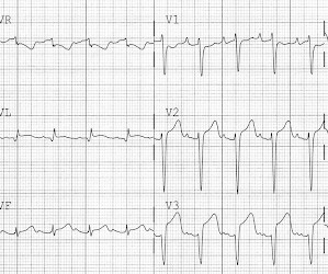
1,2 Neuroleptic malignant syndrome (NMS) (hyperthermia, autonomic instability, rigidity, altered mental status [AMS]) can occur as well and is most often seen with clozapine but has been observed with other atypicals. Rigidity and hyperthermia should raise concerns for NMS. 1 Seizures may occur due to lowered seizure threshold.


Let's personalize your content