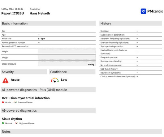
This site uses cookies to improve your experience. To help us insure we adhere to various privacy regulations, please select your country/region of residence. If you do not select a country, we will assume you are from the United States. Select your Cookie Settings or view our Privacy Policy and Terms of Use.
Cookie Settings
Cookies and similar technologies are used on this website for proper function of the website, for tracking performance analytics and for marketing purposes. We and some of our third-party providers may use cookie data for various purposes. Please review the cookie settings below and choose your preference.
Used for the proper function of the website
Used for monitoring website traffic and interactions
Cookie Settings
Cookies and similar technologies are used on this website for proper function of the website, for tracking performance analytics and for marketing purposes. We and some of our third-party providers may use cookie data for various purposes. Please review the cookie settings below and choose your preference.
Strictly Necessary: Used for the proper function of the website
Performance/Analytics: Used for monitoring website traffic and interactions
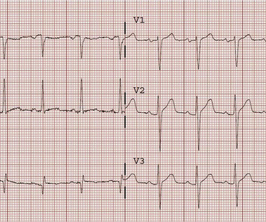
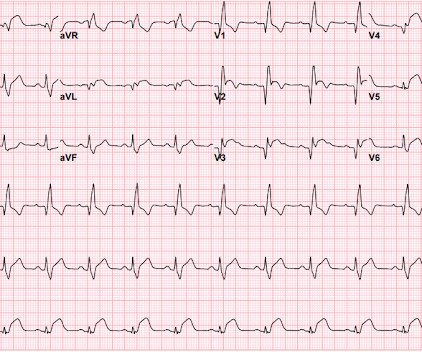
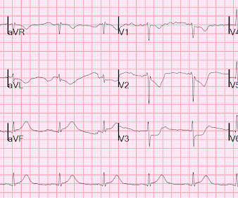
This was sent by an undergraduate (not yet in medical school, but applying now) who works as an ED technician (records all EKGs, helps with procedures, takes vital signs) and who reads this blog regularly. The undergraduate's analysis: This EKG shows J point elevation of about 0.5-1 Edited by Smith He also sent me this great case.
Pain improved to 1/10 after EMS administers 324 mg aspirin and the following EKG is obtained at triage. If this EKG were handed to you to screen from triage without any clinical information, what would you think? Do you appreciate any dynamic changes compared to the patient’s prior EKG? What do you think? In fact, Kosuge et al.
EKG from triage: Here is his previous ECG: Normal ST Elevation Resident's interpretation: Reperfusion pattern/Wellens' with biphasic T waves in V2 and V3, and in comparison to an EKG in 2020 this is new. Bedside ultrasound with no apparent wall motion abnormalities, no pericardial effusion, no right heart strain.
1 Just one year prior, Dr. Clarke had begun an emergency medicine residency at what was then known as LA CountyUSC Hospital, Los Angeles. Firsthand Account An ambulance bay at the LA County-USC Hospital in 1978. Dr. Clarke was then hired as assistant director in the emergency department (ED) at Pomona Valley Hospital, Pomona, Calif.
I was texted this ECG just as I was getting into bed. Ken (below) is appropriately worried about pulmonary embolism from the ECG. What I had not told him before he made that judgement is that the patient also had ultrasound B-lines of pulmonary edema. Figure-1: I've labeled the initial ECG in today's case.
Bedside ultrasound showed volume depletion and no pulmonary edema. Here is the prehospital ECG: First ED ECG What do you think? But I changed my mind after seeing the old ECG (below) I later sent it to Ken Grauer, who annotated as below with the red Xs: The "Y" in lead II across the bottom appears to be a PVC.
An ECG will also help with anaesthetic planning Bloods: CRP, U&E, FBC, LFTs, INR (if on warfarin), VBG (for lactate, pH and glucose), amylase Group and save: not all surgical procedures need group and saves- these are expensive and in many cases, unnecessary- check first!
Bedside ED ultrasound showed exceedingly poor global LV function, and no B lines. Here is the initial ED ECG. Other thought this was due to hyperkalemia, but the ECG does not have the appearance of hyperkalemia but does have the appearance of severe cardiomyopathy -- LBBB with very wide QRS) 3. What do you think?
The below ECG was recorded. The ECG shows obvious STEMI(+) OMI due to probable proximal LAD occlusion. This ECG does not have the typical ST-vector of an LAD occlusion. See below for Ken Grauer Comment on the initial ECG: == On arrival, another ECG was recorded: There appears to have been quite a bit of spontaneous reperfusion!
The medics recorded this prehospital ECG at 1535 : There is ST elevation and tall T-waves in precordial leads, with reasonably good R-wave progression. First, if an old ECG is available, then compare. 1.196 x STE60 in V3 in mm) + (0.059 x computerized QTc) - (0.326 x RA in V4 in mm) Third, one can do an immediate cardiac ultrasound.
A 76-year-old woman presents to a community hospital after waking with garbled speech and right-sided weakness. Case 1: Excess An elderly woman is admitted to a community hospital with a minor stroke. The hospital does an excellent job. By the time she leaves the hospital, she has no residual symptoms. I love my job.
A submassive PE does not result in hypotension but does result in right ventricular (RV) dysfunction (as evidenced by CT or ECG) and/or signs of myocardial injury, such as elevated B-type natriuretic peptide, elevated troponin, or new ECG changes. 6 The second common catheter-directed approach is mechanical thrombectomy.
Triage EKG: What do you think? He had been seen several weeks ago at an outside hospital for a similar issue and had been discharged home, presumably after unremarkable workup. A prior ECG was available for comparison. Immediately after the second ECG was performed, the patient's pain resolved completely.
There are 38 states where it currently is a felony of some level to assault a healthcare worker BUT there is currently no federal law that protects hospital employees from assault or intimidation. Current bills in progress: US H.R.2663 2663 & S.1176 1176 focuses on violence prevention (introduced for the 3rd time 4/18/23); US H.R.2584
Does that normal troponin and ECG obviate the need for cardiology consultation for my patient with a concerning story for acute coronary syndrome? The “bundling” heuristic—if I send a troponin, then an ECG is needed. A testicular ultrasound confirmed restored blood flow. Mary’s Hospital in Leonardtown, Maryland.
Initial ROSC was obtained, during which this ECG was obtained: What do you think? I sent it to 2 of my ECG nerd colleagues with no clinical information whatsoever, who instantly said: "Looks like afib with subendocardial ischemia and right heart strain pattern." "I CPR was initiated immediately. The rhythm is atrial fibrillation.
Angiography after Out-of-Hospital Cardiac Arrest without ST-Segment Elevation. Angiography after Out-of-Hospital Cardiac Arrest without ST-Segment Elevation. The EKG shows sinus tachycardia with nonspecific changes and no ST segment elevations, Q waves, or hyperacute T waves. Date: September 8th, 2021 Reference: Desch et al.
She has no previous cardiac history of which she is aware 911 was called and here is her Prehospital ECG: What do you think? link] Case continued She arrived in the ED and here is the first ED ECG. In comparison with the first ECG, I would guess that the artery is reperfusing. I don't know if her pain was getting better or not.
The ECG told the story. EMS recorded these prehospital ECGs: Time 0: In V2-V4, there is ST elevation that does not meet STEMI "criteria," of 1.5 Time = 24 min S-wave depth is diminishing These prehospital ECGs were lost and not seen. This patient has a non-diagnostic ECG by most rules. She called 911. STE 60 V3 = 1.5
1-3 VTE is responsible for hospitalizing over 250,000 Americans every year, and there are an estimated 100,000 deaths annually associated with these conditions. 1-3 VTE is responsible for hospitalizing over 250,000 Americans every year, and there are an estimated 100,000 deaths annually associated with these conditions. EKG RV strain.
This case was provided by Spencer Schwartz, an outstanding paramedic at Hennepin EMS who is on Hennepin EMS's specialized "P3" team, a team that receives extra training in advanced procedures such as RSI, thoracostomy, vasopressors, and prehospital ultrasound. Our first 12 lead ECG was then recorded at 0926. What do you think?
If you want to see Justin in the video format and his new Youtube series then click here: [link] If you would like to check out my current ultrasound training project then click here: WACHS Ultrasound Otherwise – enjoy the show and see you in a few weeks. Myth 3: The ECG changes from hyperkalemia are predictable and reliable.
This is her pre-hospitalECG: This is her first ECG in the ED: What do you think? However, the prehospital ECG is more worrisome: the T-wave inversion in V5 and V6 is preceded by ST Elevation. When I say " relative ," I mean relative to the previous ECG, which is the baseline, chronic, non-ischemic ECG.
This case was sent by Dr Avinash Krishnamurthy, a fine emergency medicine resident from Australia Cairns base hospital Case : An adolescent male had a mechanical fall and injured his left shoulder and arm. A bedside cardiac ultrasound was normal. An ECG was recorded: Avinash was understandably confused by this ECG.
ECG: Evaluate for ischemia, right heart strain. Ultrasound Sensitivity 88-100%, specificity 68-94% Positive likelihood ratio of 14.6 (95% Consider risk factors for multi-drug resistant microbes: Recent IV antibiotics Hospitalization within 90 days. Overestimates arterial oxygen saturation. Consider CT with IV contrast.
Past medical history included RBBB without other cardiac history, but old ECG was not available. As for the ECG, it could represent OMI, but RBBB is also a clue that it may be PE. But with prehospital and ED ECGs being ‘STEMI negative’, the ECG was signed off and the patient waited to be seen.
ECG, CXR, and troponin are negative. Troponins #1 and #2 are borderline and ECG is non-specific. In the ED, his troponin, ECG, and chest X-ray (CXR) are normal. ECG, CXR, and troponin are all normal. An ECG and CXR are negative. An ECG, CXR, and two troponins are normal. She is given morphine for pain.
The patient needs a transvaginal ultrasound w/ doppler with gynecology consult and possible laparotomy TVUS is not just for torsion, it can also be used for ectopic pregnancies or pregnancy of unknown location Acute Ischemic Stroke Updates WITH Drs.
ECG: Evaluate for ischemia, right heart strain. Ultrasound Sensitivity 88-100%, specificity 68-94% LR+ of 14.6 (95% Consider risk factors for multi-drug resistant microbes: Recent IV antibiotics Hospitalization within 90 days. COVID-19 and ACS COVID increases the risk of hospitalization, pneumonia, pain, and ACS in SCD patients.
The screening physician ordered an EKG and noted his ashen appearance and moderate distress. Triage EKG: What do you think? Close up of V3 with baseline in red Close up of V4 with baseline in red This ECG alone is highly suspicious for posterior OMI! This is a very specific marker of posterior OMI. Epub 2021 Nov 15.
An immediate 12-lead EKG was obtained: There is ST elevation in leads aVR and V1, with marked ST depression in I, II, III, aVF, V3-V6. Smith comment: This patient did not have a bedside ultrasound. The ECG cannot diagnose the etiology of ischemia; it only the presence of ischemia, from whatever etiology. What should be done?
A prehospital ECG was recorded: Limb leads: Precordial Leads What is the therapy? On arrival, the patient was in shock, was intubated, and had an immediate cardiac ultrasound. What does a heart look like on ultrasound when the EKG looks like that? The followup ECG is here: Now the QRS is only slightly prolonged.
Note 2 other similar cases at the bottom that come from my book, The ECG in Acute MI. Case While I was busy seeing patients, a resident brought me this ECG of a 60-something with a history of syncope only. There was no chest pain or SOB at the tim of the ECG: Computerized QTc is 464 ms A previous ECG from 8 years prior was normal.
Myth: Some patients are “too sick to transport” Myth Part B: The receiving hospital gets to decide if a patient is too sick to transport. If you or your patient do not have a watch capable of providing you with an ECG, consider asking those on board to allow you to borrow theirs. What drives the concern?
He had the following EKG recorded: Low voltage, suggests effusion. A bedside cardiac ultrasound was normal, with no effusion. He was started on Extracorporeal Life Support ("VA ECMO") Here is the ECG on ECMO: Very low voltage On Day 3, the EF recovered (that seems quick!) He was moderately hypoxic. 3–8 Shi et al.
In select patient populations (such as those with poor outpatient follow-up), screening with a BMP to evaluate their serum creatinine may be beneficial Patients with asymptomatic hypertension should be referred to their PCP for outpatient follow-up.
The paramedics found the patient with ROSC and a GCS 7, and an ECG showing LBBB with possible lateral ST elevation. Below is the first ED ECG, labeled LBBB by the machine. Similarly, STEMI guidelines call for urgent angiography for refractory ischemia or electrical/hemodynamic instability, regardless of ECG findings.
Here is her ECG on arrival to the ED: What is your differential? The ECG is quickly reviewed and shows sinus rhythm with normal QRS complexes. Her prior ECG on file is shown below: What are your next steps? Learning Points: The ECG is always just one piece of the clinical puzzle. What are your next steps?
It is unclear whether he had active pain at the time of the first ECG: What do you think? 0.20 (above which, the ECG likely represents OMI). The team was concerned about possibly ischemic ST elevations on the ECG above, so they took it to Dr. Nossen for consultation. Vitals were within normal limits.
Her ECG did not show any concerning features. The patient was referred to hospital to further investigate the cause of her collapse, in light of the uncertain aetiology of the echo findings. HH has been associated with post-prandial syncope, chest pain, and ECG changes suggestive of ischaemia. Cardiovascular ultrasound.
What is your ECG interpretation and what would you do next? This ECG shows a normal sinus rhythm with a normal conduction pattern (normal PR, normal QRS, and normal QTc), normal axis, late R wave progression (and misplaced V2), normal voltages, ST-elevation in aVR and global ST-depressions. BP was 110 and oxygen saturation was normal.
Meyers ECG Interpretation: Easily diagnostic of LAD occlusion. The ECG easily meets STEMI criteria in all leads V2-V6, as well. I sent this ECG to Dr. Smith, with the only information that it is a 17 year old with chest pain. The ECG was interpreted as "benign early repolarization." Coxsackie serologies negative.
Bedside ultrasound showed no effusion and moderately decreased LV function, with B-lines of pulmonary edema. Here is his ED ECG: There is obvious infero-posterior STEMI. This subsequent ECG was recorded after the K was up to 2.2 These two rhythms are often indistinguishable on the monitor or ECG. This was stented.
When the paramedics arrived, they obtained a 12 lead ECG and confirmed the unstable vital signs. EKG is pictured below: What do you think? They did not have an ultrasound on the ambulance (some local crews are starting to utilize POC limited US in our service areas). A right sided ECG was not recorded.
We organize all of the trending information in your field so you don't have to. Join 5,000+ users and stay up to date on the latest articles your peers are reading.
You know about us, now we want to get to know you!
Let's personalize your content
Let's get even more personalized
We recognize your account from another site in our network, please click 'Send Email' below to continue with verifying your account and setting a password.
Let's personalize your content