Remove EKG/ECG Remove Fluid Resuscitation Remove Wellness
article thumbnail

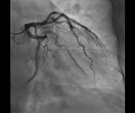
Anterior OMI. What does the angiogram show?

Dr. Smith's ECG Blog

His ECG is shown below. The LCx and OM branches are not as well seen. How does RCA occlusion fit with the ECG? The answer is that the ECG really shows right ventricular OMI. Written by Willy Frick A 50 year old man with no medical history presented with acute onset substernal chest pain.

EKG/ECG 107
article thumbnail

Tachycardia must make you doubt an ACS or STEMI diagnosis; put it all in clinical context

Dr. Smith's ECG Blog

He had this ECG recorded. He was rushed by residents into our critical care room with a diagnosis of STEMI, and they handed me this ECG: There is sinus tachycardia with ST elevation in II, III, and aVF, as well as V4-V6. But, remember, we do not evaluate and treat ECGs, we evaluate and treat patients. Are the lungs clear?

EKG/ECG 52
Insiders

Sign Up for our Newsletter

This site is protected by reCAPTCHA and the Google Privacy Policy and Terms of Service apply.

Trending Sources

article thumbnail

EM@3AM: Amniotic Fluid Embolism

EMDocs

The newborn is doing well, but the mother is complaining of shortness of breath and chest pain. A 37-year-old G5P4 at 33 weeks presents to the ED after being brought in by ambulance. She had a precipitous delivery while the ambulance was pulling in. Triage vital signs (VS) include BP 88/45, HR 121, T 97.1, RR 28, SpO2 89% on 6L NC.

EMS 96
article thumbnail

Electrical injuries

Don't Forget the Bubbles

Suspected low voltage injuries (<1000v) should still be approached with an A-E assessment, but if 12 lead ECG and urinalysis are normal, it’s reasonable to discharge the patient. If the ECG is abnormal, the patient should be managed as if exposed to a high voltage (>1000v).

Burns 80
article thumbnail

Diffuse Subendocardial Ischemia on the ECG. Left main? 3-vessel disease? No!

Dr. Smith's ECG Blog

An immediate 12-lead EKG was obtained: There is ST elevation in leads aVR and V1, with marked ST depression in I, II, III, aVF, V3-V6. In addition, the patient received 750 mL of fluid resuscitation with transient improvement of blood pressure. What should be done? Should the cath lab be activated?

EKG/ECG 40
article thumbnail

Putting Clinical Gestalt to Work in the Emergency Department

ACEP Now

In such cases, would you wait for a lactate, white blood cell count, bandemia, or other diagnostics to confirm a source of infection before starting antibiotics, fluid resuscitation, and/or pressors? In this study, clinical gestalt is not only fast, but accurate for the benefit of timely resuscitation and intervention.

article thumbnail

Chemical Burns

Mind The Bleep

Alkali burns result in liquefaction necrosis, allowing for deeper tissue injury as well as vascular injury that can lead to both local and systemic toxicity [1]. Circulation Assess heart rate, blood pressure, peripheral and central CRT, pulses and 3 lead ECG. Establish IV access and begin fluid resuscitation with 250ml boluses of 0.9%

Burns 52