Remove EKG/ECG Remove Fluid Resuscitation Remove Resuscitation
article thumbnail

Diabetic Ketoacidosis in Paediatrics

Mind The Bleep

ECG: to monitor T wave changes due to hypokalaemia. ECG features of Hypokalaemia: Increased P wave amplitude (peaked P waves) Prolonged PR interval Widespread ST depression T wave flattening or inversion Prominent U waves (most noticeable in the precordial leads) Figure 2 : ECG of a patient with serum K+ of 1.9

article thumbnail

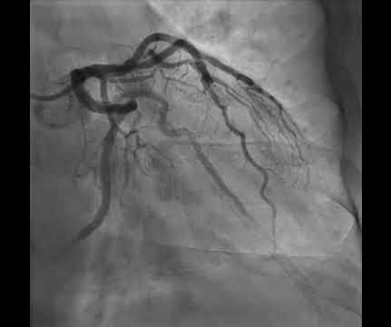
Anterior OMI. What does the angiogram show?

Dr. Smith's ECG Blog

His ECG is shown below. How does RCA occlusion fit with the ECG? The answer is that the ECG really shows right ventricular OMI. We've highlighted a considerable number of acute RV MI cases in Dr. Smith's ECG Blog ( See the October 7, 2019 and May 10, 2024 posts , to name just two ). There is non-obstructive disease here.

EKG/ECG 110
Insiders

Sign Up for our Newsletter

This site is protected by reCAPTCHA and the Google Privacy Policy and Terms of Service apply.

article thumbnail

EM@3AM: Amniotic Fluid Embolism

EMDocs

Answer : Amniotic fluid embolism Epidemiology: Incidence of 1:15,200 to 1:53,400 1 7% occur during labor Causes approximately 14% of all maternal peripartum death in United States Current fatality rate 13-60% 1-4 Risk factors: Advanced maternal age, amniocentesis, cesarean delivery, eclampsia, medical induction of labor, placental pathology, diabetes, (..)

EMS 97
article thumbnail

Electrical injuries

Don't Forget the Bubbles

Suspected low voltage injuries (<1000v) should still be approached with an A-E assessment, but if 12 lead ECG and urinalysis are normal, it’s reasonable to discharge the patient. If the ECG is abnormal, the patient should be managed as if exposed to a high voltage (>1000v). Resuscitation. Culnan, D.M., Jimenez, C.

Burns 80
article thumbnail

Tachycardia must make you doubt an ACS or STEMI diagnosis; put it all in clinical context

Dr. Smith's ECG Blog

He had this ECG recorded. He was rushed by residents into our critical care room with a diagnosis of STEMI, and they handed me this ECG: There is sinus tachycardia with ST elevation in II, III, and aVF, as well as V4-V6. But, remember, we do not evaluate and treat ECGs, we evaluate and treat patients. The K returned at 6.9

EKG/ECG 52
article thumbnail

CICM Second Part Exam Practice SAQs 22082024

Intensive Blog

Outline the initial assessment of severe burns injury, including how you will calculate fluid resuscitation requirements. cerebral ox, ECG, TOE, differential BPs) Bleeding? Discuss the advantages and disadvantages of using pulse oximetry to target oxygen therapy (50%) What SpO2 targets do you use in ICU patients and why? (50%)

Burns 52
article thumbnail

emDOCs Podcast – Episode 100: Acute Chest Syndrome Part 1

EMDocs

ECG: Evaluate for ischemia, right heart strain. Fluid management Goal is euvolemia Dehydration – needs IV fluid resuscitation. Hypervolemia – leads to pulmonary edema: Consider diuresis If euvolemic – start maintenance fluids of D5 in 0.45%NS Overestimates arterial oxygen saturation.