article thumbnail

CICM Second Part Exam Practice SAQs 22082024

Intensive Blog

Outline the initial assessment of severe burns injury, including how you will calculate fluid resuscitation requirements. cerebral ox, ECG, TOE, differential BPs) Bleeding? Discuss the advantages and disadvantages of using pulse oximetry to target oxygen therapy (50%) What SpO2 targets do you use in ICU patients and why? (50%)

Burns 52
article thumbnail

The Intersections of Physical and Mental Health Disorders

ACEP Now

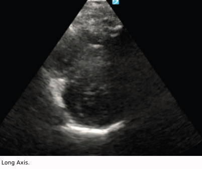
However, the persistence of tachycardia despite fluid resuscitation and dosing with lorazepam to help with his panic disorder raised concerns that warranted further investigation. Our workup started with an ultrasound and EKG. EKGs will have abnormalities in about 90 percent, but they are typically nonspecific findings.

EKG/ECG 52
article thumbnail

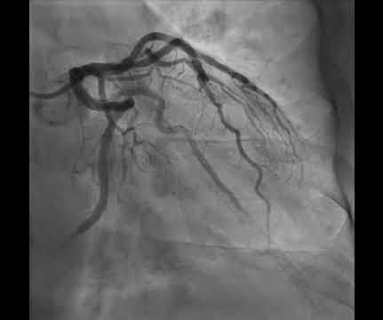
Anterior OMI. What does the angiogram show?

Dr. Smith's ECG Blog

His ECG is shown below. How does RCA occlusion fit with the ECG? The answer is that the ECG really shows right ventricular OMI. We've highlighted a considerable number of acute RV MI cases in Dr. Smith's ECG Blog ( See the October 7, 2019 and May 10, 2024 posts , to name just two ). There is non-obstructive disease here.

EKG/ECG 109
article thumbnail

Electrical injuries

Don't Forget the Bubbles

Suspected low voltage injuries (<1000v) should still be approached with an A-E assessment, but if 12 lead ECG and urinalysis are normal, it’s reasonable to discharge the patient. If the ECG is abnormal, the patient should be managed as if exposed to a high voltage (>1000v).

Burns 80
article thumbnail

Diabetic Ketoacidosis in Paediatrics

Mind The Bleep

ECG: to monitor T wave changes due to hypokalaemia. ECG features of Hypokalaemia: Increased P wave amplitude (peaked P waves) Prolonged PR interval Widespread ST depression T wave flattening or inversion Prominent U waves (most noticeable in the precordial leads) Figure 2 : ECG of a patient with serum K+ of 1.9

article thumbnail

Another deadly and confusing ECG. Are you still one of the many people who will be fooled by this ECG, or do you recognize it instantly?

Dr. Smith's ECG Blog

Fluid resuscitation was initiated. Here is the initial ED ECG: What do you think? The ECG shows peaked T waves, QRS widening, a coved-type ST segment with inverted T waves most prominent in V1 and V2 consistent with Brugada pattern. They opined that the ECG could instead be "metabolic." Creatinine elevated at 3.09

EKG/ECG 52
article thumbnail

Chemical Burns

Mind The Bleep

Circulation Assess heart rate, blood pressure, peripheral and central CRT, pulses and 3 lead ECG. Establish IV access and begin fluid resuscitation with 250ml boluses of 0.9% This includes adequate pain control, fluid resuscitation, and stabilization of any systemic complications.

Burns 52