Remove EKG/ECG Remove EMS Remove Wellness
article thumbnail

ECG Blog #419 — The Cause of ECG #1?

Ken Grauer, MD

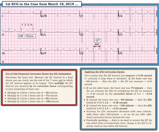
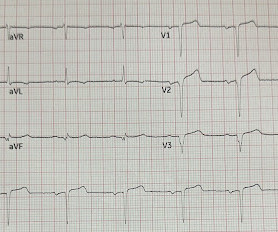
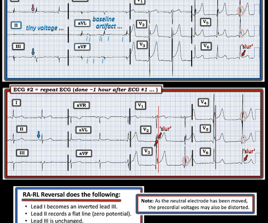
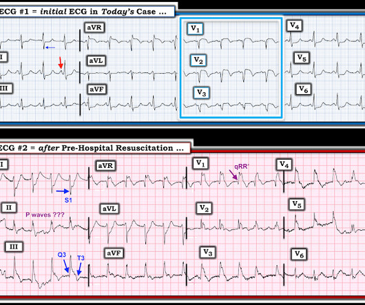
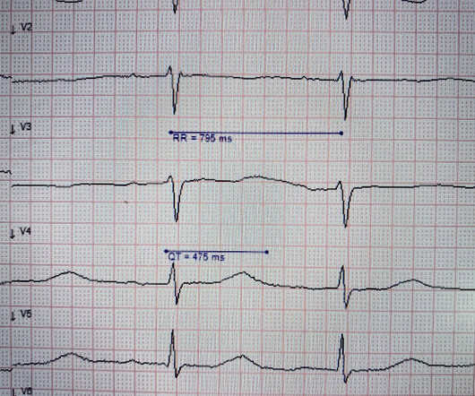
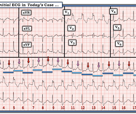
I was sent the 2 ECGs shown in Figure-1 — which were recorded from an elderly man whose heart beat "has been irregular for years". No clear history for recent chest pain — but the patient "has not been well" for the previous week. QUESTIONS: How would YOU interpret these 2 ECGs? — How might ECG #2 be related to ECG #1 ?

EKG/ECG 496
article thumbnail

ECG Blog #406 — To Do Additional Leads?

Ken Grauer, MD

For full discussion of this case — See ECG Blog #351 — == The ECG in Figure-1 — was obtained from a previously healthy older man who contacted EMS ( E mergency M edical S ervices ) because of "chest tightness" that began ~1 hour earlier. Given this history: QUESTIONS: How would YOU interpret the ECG in Figure-1 ?

EKG/ECG 413
Insiders

Sign Up for our Newsletter

This site is protected by reCAPTCHA and the Google Privacy Policy and Terms of Service apply.

Trending Sources

article thumbnail

ECG Blog #427 — To Cath this Elderly Patient?

Ken Grauer, MD

The ECG in Figure-1 — was obtained on the scene by EMS ( E mergency M edical S ervices ). He was hemodynamically stable — but clearly distressed with a sense of “impending doom” at the time ECG #1 was recorded. QUESTIONS: How would YOU interpret the initial ECG in Figure-1 ? Figure-1: The initial ECG in today's case.

EKG/ECG 387
article thumbnail

ECG Pointers: WPW doesn’t usually cause a problem, right?

EMDocs

This week, we feature a post from Dr. Tannenbaums ECG Teaching Cases , a free ECG resource. Without further ado, lets look at some ECGs! I dont think this is normal the tech tells you as he hands you an ECG. Hes stable for the moment, so lets take a look at his ECG: Rate: really fast! Please check it out.

EKG/ECG 67
article thumbnail

Torsade in a patient with left bundle branch block: is there a long QT? (And: Left Bundle Pacing).

Dr. Smith's ECG Blog

She had an ECG recorded: This is left bundle branch block (LBBB), with appropriate proportional discordance. Dodd KW, Elm KD, Dodd EM, Smith SW. Here is one of the strips This is clearly polymorphic VT and probably torsade de pointes Subsequent ECGs. CT of the chest showed no pulmonary embolism but bibasilar infiltrates.

EKG/ECG 116
article thumbnail

An undergraduate who is an EKG tech sees something. The computer calls it completely normal. How about the physicians?

Dr. Smith's ECG Blog

This was sent by an undergraduate (not yet in medical school, but applying now) who works as an ED technician (records all EKGs, helps with procedures, takes vital signs) and who reads this blog regularly. He called EMS, who arrived on scene about two hours after the onset of pain to find him hypertensive at 220 systolic.

EKG/ECG 126
article thumbnail

Concerning EKG with a Non-obstructive angiogram. What happened?

Dr. Smith's ECG Blog

EMS obtained the following vital signs: pulse 50, respiratory rate 16, blood pressure 96/49. It appears EMS obtained two EKGs, but unfortunately these were not saved in the medical record. The EMS crew was only BLS certified, so EKG interpretation is not within their scope of practice.

EKG/ECG 127