CONCEALED CONDUCTION
ECG Guru
JANUARY 12, 2025
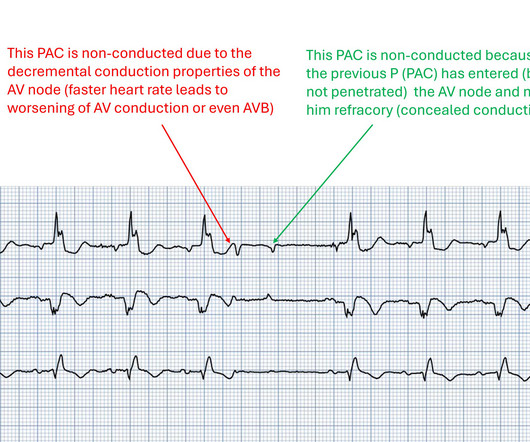
This ECG (3 rhythm strips) initially shows a sinus rhythm with 1st degree AVB grade I and wide QRS complexes (presumably RBBB pattern). A PAC (P-wave premature, different form than in sinus rhythm) appears approximately in the middle of the ECG, this is not conducte. Due to the strong prematurity, this is not surprising. But why is the 2nd PAC also blocked?


Let's personalize your content