Remove Documentation/Coding Remove Sepsis Remove Ultrasounds
article thumbnail

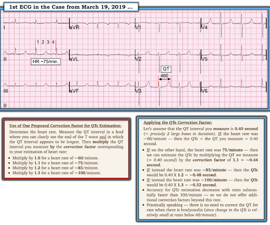
Torsade in a patient with left bundle branch block: is there a long QT? (And: Left Bundle Pacing).

Dr. Smith's ECG Blog

Bedside cardiac ultrasound showed moderately decreased LV function. She was managed for sepsis with antibiotics including azithromycin, had hypotension with arterial and central lines placed and pressors. , but potassium returned normal. In the middle of the night, a "code" was called, and multiple rhythms like this were recorded.

EKG/ECG 115
article thumbnail

Putting Clinical Gestalt to Work in the Emergency Department

ACEP Now

Our nurse did not study Paul Ekman’s Facial Action Coding System for Action Units to code “fear” in the patient’s face. This means deviating from protocols (when warranted) for sepsis because your initial impression is the patient would be harmed with that much fluid. A testicular ultrasound confirmed restored blood flow.

Insiders

Sign Up for our Newsletter

This site is protected by reCAPTCHA and the Google Privacy Policy and Terms of Service apply.

Trending Sources

article thumbnail

emDOCs Podcast – Episode 98: Post ROSC Mental Model

EMDocs

Check the pulse RSI= Resuscitation Sequence Intubation Hypoxia, Hypotension, and Acidosis are the reason patients code during/post intubation These patients are super high risk for all 4 Optimize first pass success – Induction agent + paralytic Unconscious patients will still have muscle tone Induction Ketamine or Etomidate at half doses (i.e.,

article thumbnail

The Latest in Critical Care, 2/5/24 (Issue #28)

PulmCCM

You can read the document here. Read the original document here. In 18 severe sepsis patients in 3 years at one center who underwent PET scanning (apparently done ad hoc), 14 of the 18 had positive tests, and 11 were true positives, leading to surgery in 2 and pacemaker removal in 2 (and longer / new antibiotics in 2).

article thumbnail

Diagnostics and Therapeutics: The Who, What, Where, When and Why of Lumbar Punctures

Taming the SRU

1) However, it wasn’t until the 1890s that purposeful, successful, and safe attempts to access this fluid were documented (2). It is most helpful to do the ultrasound immediately before needle insertion, as movement of the patient may shift cutaneous landmarks from underlying bony structures. WHY - Why Not? REFERENCES 1.

article thumbnail

Diffuse Subendocardial Ischemia on the ECG. Left main? 3-vessel disease? No!

Dr. Smith's ECG Blog

Smith comment: This patient did not have a bedside ultrasound. Had one been done, it would have shown a feature that is apparent on this ultrasound (however, this patient's LV function would not be as good as in this clip): This is recorded with the LV on the right. In fact, bedside ultrasound might even find severe aortic stenosis.

EKG/ECG 40
article thumbnail

Episode 30 - Emergency Department Management of Patients With Complications of Bariatric Surgery

EB Medicine

Leakage from the staple line typically presents within the first week, but can present up to 35 days, usually with fevers, tachycardia, abdominal pain, nausea, vomiting sepsis, or peritonitis. Some may present with isolated tachycardia while others may present with profound sepsis – tachycardia, hypotension, and fever. At 6 to 8 mL/kg.