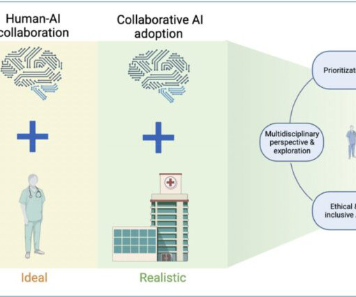
This site uses cookies to improve your experience. To help us insure we adhere to various privacy regulations, please select your country/region of residence. If you do not select a country, we will assume you are from the United States. Select your Cookie Settings or view our Privacy Policy and Terms of Use.
Cookie Settings
Cookies and similar technologies are used on this website for proper function of the website, for tracking performance analytics and for marketing purposes. We and some of our third-party providers may use cookie data for various purposes. Please review the cookie settings below and choose your preference.
Used for the proper function of the website
Used for monitoring website traffic and interactions
Cookie Settings
Cookies and similar technologies are used on this website for proper function of the website, for tracking performance analytics and for marketing purposes. We and some of our third-party providers may use cookie data for various purposes. Please review the cookie settings below and choose your preference.
Strictly Necessary: Used for the proper function of the website
Performance/Analytics: Used for monitoring website traffic and interactions
Chuck Pilcher, MD, FACEP Editor, Medical Malpractice Insights Editor, Med Mal Insights Radiology over-reads – Who’s responsible? Patient not informed of enlarged heart, dies 3 weeks post ED visit Miscommunicated radiology findings are a hot topic. This patient’s history was either poorly taken or poorly documented.
The authors also tried to evaluate the frequency and outcomes of laboratory and radiology tests ordered by emergency physicians. There are going to be issues with confounding, mis-coded data, and variation across sites. Everything here is almost assuredly imprecise and unable to be generalized outside the VA system involved.
Don’t panic: If you are the only available person and dont know the patient, document the information in the notes and let your senior know. Again, document the call clearly, discuss with your senior and implement further investigations/treatment as appropriate. link] Written by Emma Chrisholm ST1 Radiology
Either they should ask the surgical team for clinical management of a patient with suspected septic ileus with a differential including necrotising enterocolitis or the radiology team for interpretation of the x-ray. What to do when referrals are not accepted Ask “Why?”
They separated all patients with acetabular and pelvic ring fractures using ICD-10 codes. If there is concern that there could be ongoing bleeding, PPP can be followed by a trip to the interventional radiology suite. This eliminated patients who might have received other additional management that could cloud the data.
Does this patient require radiologic investigations? The clinician reviewers – one resident physician and one attending physician – did not much agree (73-83% agreement) on admission, radiology, and antibiotic determinations. Specifically, the LLM – here GPT-3.5T and GPT-4T – is asked: Should this patient be admitted to the hospital?
.” The tech cleans the wound and documents it as 2cm long and “ dirty.” The ED physician documents suturing a 3 cm laceration in a single layer, omitting any reference to any wound exploration, cleansing or debridement. I thought they were incidental “ bone islands” so I didn ’ t document them. Negligence was present.
These multiple hazards include biological, chemical, ergonomic, and radiological risks. This can be achieved by maintaining a centralized document repository. Have a digital document management system or a physical binder in a prominent area of the dental office.
A typical shift when he was starting out would include patients falling into what was coded as 1350 major medical/trauma, 1060 minor medical/trauma, or 1050 medical walk-in. The advancement of radiology was the first thing that came to mind for Dr. Clarke when discussing how technology has advanced emergency medicine.
This fills in the code on the second tab which will be added to TraumaBase. If you use a different registry, contact your vendor for assistance. The spreadsheets consist of three tabs/pages. On the first, enter the specific field names from your TraumaBase implementation.
If we include PACS with the EMR, there was also the waiting at the radiology file room window, while a clerk tried to find an old X-ray. ” We repeated the CT, which revealed bilateral pulmonary emboli, and recalled the days when the radiologist would have instructed about the inadequacy of the scan during post-call radiology rounds.
Exam is normal except for tenderness as documented in the diagram. She never had a documented abdominal exam. I just failed to document it. The EP documented seeing the patient and agreed with the ARNP’s plan. If you take a history or examine a body part, document it. Temp is 98.7 and pulse 121.
13,16 In cases of radiological or nuclear incidents, follow referenced contamination and exposure protocols, including promptly consulting the radiation safety officer, wearing protective equipment, initiating decontamination measures and evaluating for acute radiation syndrome if indicated. J Appl Clin Med Phys. 2022;23(3):e13526.
Method and Design The ED and radiology departments collaborated to develop an MRI direct to scan (DTS) pathway bypassing radiology discussions. All DTS scans had documented senior review at radiology request to ensure governance. There has been an increase in number of positive cases from 4.2% to 6.5%.
Document a risk and benefit discussion with the patient regarding empiric anticoagulation, perhaps even including the HAS-BLED score in the documentation [10]. Thrombosis in the emergency department: use of a clinical diagnosis model to safely avoid the need for urgent radiological investigation. mg/kg for 24 hours.
I have been a part of many traumatic codes and resuscitations during my training, but I knew this one would probably be my most difficult one. The radiology tech has his hand on a weak femoral pulse. Should be here in 5 minutes.” reported one of the seasoned nurses. She was calm. My response: “Call everyone. Prepare everything.
” However, when we combine POCUS images with clinical documentation, we are telling a story — and every good story has a beginning, middle, and end. Point-of-care ultrasound (POCUS) is often taught to answer binary questions, like, “Is there free fluid in the abdomen?”
ultrasound grand rounds: bedside dvt studies - family presence in the ed/icu - r1 clinical knowledge: aicd - r3 small groups: difficult airway management Ultrasound grand rounds: DVT studies WITH Dr. minges Why should we perform bedside DVT studies in the ED? ETT onto a fiberoptic scope.
She presents with a folder of records and codes for outside EMRs. It is when you diagnose amyloidosis as the cause of a peripheral neuropathy, or find that the cause of a patient’s anxiety is hyponatremia, caused by SIADH, caused by a radiologically undetectable lung cancer. Sensible Medicine is a reader-supported publication.
Craniosynostosis, The Radiology Assistant. The Radiology Assistant: Craniosynostosis Accessed 10.01.2024 Mawji, A., Recommendations for sudden infant death syndrome prevention: a discussion document. Prevalence, risk factors, and natural history of positional plagiocephaly: a systematic review. Vollman, A. Hatfield, J.,
vs. It is worth mentioning that this study was a multicenter study based in the US with representation from ED, Radiology, and Urology. Third, for those with proper training, and with some exceptions (see the systematic review paper for case vignettes that highlight these), POCUS is non-inferior to radiology-performed US.
Surgery was completed approximately one month after presentation with improvement in vision to 20/80 at last documented ophthalmology follow up appointment. Ectopia Lentis: Radiology Reference Article.” OMFS was consulted for multiple facial fractures for which patient was set up to follow up in clinic. Cureus , Cureus, 24 Apr.
Pneumonia diagnosis supported by clinical and radiologic criteria. While patients received respiratory support, supportive care, and antibiotics at the discretion of the medical team, the specific interventions provided outside the study were not documented, potentially leading to co-intervention bias.
It is worth looking through previous documentation to see whether the patient’s team or senior intensivists have set any individual cardiovascular targets. Secretion burden and weak cough are common reasons for failed extubations, so this is a vital piece of information to guide clinical decisions.
A chest X-ray was obtained: This was read by radiology as "Bilateral lower lobe interstitial opacities. We should not have been fooled by the radiology reading of "pneumonia". This was not recognized. The patient was started on a nitro drip. The initial troponin I returned at 0.67 ng/mL (URL = 0.030 ng/mL).
CT is good but you really should learn ultrasound, and lastly, sick patients need prompt consultation and resuscitation, not rapid trips to radiology. Unstable patients do not belong in a radiology suite - they belong in the ED resus bay to be resuscitated first. Nachi: All valid points, but let’s dive in too some actual detail.
Generally accepted (and some airlines spell it out) that they are assuming the liability even for you helping Documentation: To protect yourself (and the airline), will/should make documentation of what happened. Airline companies have been sued. Each airline generally has their own protocol.
The best documented cormorbidity is sickle cell disease, although other pro-thrombotic conditions also put the child at risk. Myocardial infarction in an adult resulting from coronary aneurysms previously documented in childhood after an acute episode of Kawasaki’s disease. 1986; 108(2):198-203. Pongratz G et al. European Heart J.
Rebuilding of EDs in proximity to Radiology, Blood Bank, OR, and ICU, becoming organic to the hospital. EMS in America: The Foundation Documents. Milestones of Modern Progress in Emergency Care: WWII and Korean War military medical experience and progress. 1950s & 1960s Resuscitation Research. Touchstone, Mike. Sakran, J.
Vitamin D, B12, Calcium, foate, iron, and thiamine deficiencies are all well documented complications. PA’s and NP’s - make sure to use the code APP4 at checkout to save 50%. Internal hernia after laparoscopic Roux-en-Y gastric bypass: a correlation between radiological and operative findings. J Gastrointest Surg.
Posterior – Shortened, adducted, internally rotated 5,6 Anterior – Shortened, abducted, externally rotated 5,6 As with all dislocations, perform and document distal neurovascular exam before and after management. 8 Perform and document distal neurovascular exam. 5 CT if concern for occult fracture.
This study relied on coding, and if the patient had not been coded as asthma, for example, and coded as pneumonia instead, these patients would not have been included in the study set. This highlights the need for departments to ensure timely radiology reports and continuous education of HCPs. and 16.7% , respectively.
Outcomes: Primary: Percent change in management decisions after introducing the Radiology Department RUQ US. A change in clinical course resulting from a discrepancy between POCUS and radiology department imaging only occurred in 2% of patients (not 10%).
Clinical and radiological findings supported the diagnosis of pneumonia. A QIP performed in a paediatric intensive care unit aimed to reduce unnecessary LFT requests by implementing a blood test request form and a table of common investigations to facilitate and document discussion between the nursing and medical teams.
This neurological evaluation should include assessment and documentation of the GCS, the presence of any neurologic deficits, and an NIHSS. 10 Additionally, discussion with radiology and neurology is advisable prior to obtaining an MRI, as data is not available to guide the specific timing of the test following symptom onset.
Jeff: All patients with a positive pregnancy test and vaginal bleeding should receive an ultrasound performed by either an emergency physician or by radiology. For a completed abortion, you would expect a closed OS with no IUP on ultrasound with a previously documented IUP.
mg/kg of IBW Seek to match a patient's minute ventilation with TV and RR after intubation Intentionally match patient's intrinsic RR noted prior to intubation Note that healthy lungs can handle 8 mL/kg, based on IBW, if that is useful for compensation Secure your ETT Thomas Tube Holder is a new ETT holder on Air Care for ETT down to size 6.5
A nurse documents soft tissue tenderness in the back; the EPs note describes no tenderness and only decreased ROM of the back. The radiology report even advised If this study fails to adequately explain patients symptoms, additional imaging may be indicated. On arrival she has 9/10 right lower lumbar pain. A UA is normal with no RBCs.
As mentioned, AI has tremendous potential for automating non-teaming tasks such as documentation and data retrieval, and for managing ED workflow processes. AI emergency radiology tools can lead to immediate results for high-acuity patients, reducing delay in initiating treatments for acute patients. Artif Intell Med. 2020;102:101762.
Descriptive Terms When describing injuries of the hand for documentation or referral purposes it is important to know the terminology that is widely in use in order to convey an accurate description to others. Hand dominance, mechanism of injury and vascular, neurological, and musculoskeletal status should all be evaluated and documented.
We organize all of the trending information in your field so you don't have to. Join 5,000+ users and stay up to date on the latest articles your peers are reading.
You know about us, now we want to get to know you!
Let's personalize your content
Let's get even more personalized
We recognize your account from another site in our network, please click 'Send Email' below to continue with verifying your account and setting a password.
Let's personalize your content