Remove Documentation/Coding Remove EKG/ECG Remove Wellness
article thumbnail

ECG Blog #435 — Did Cath Show Acute Ischemia?

Ken Grauer, MD

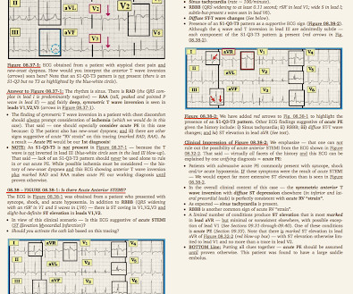
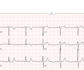
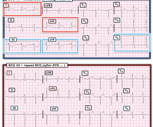
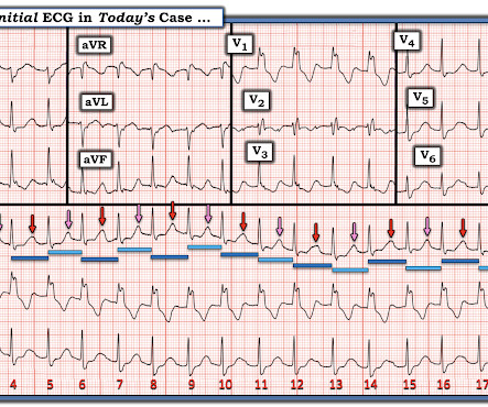
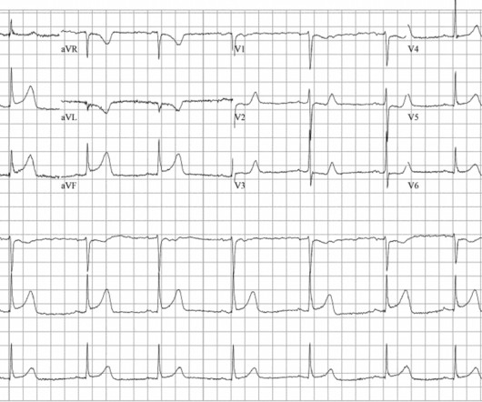
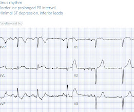
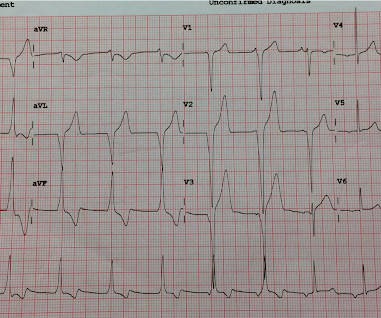
The ECG in Figure-1 — was obtained from a middle-aged woman with positional tachycardia and diaphoresis with change of position from suprine to sitting. Although CP ( C hest P ain ) was not a prominent symptom — ACS ( A cute C oronary S yndrome ) was suspected from the chest lead T wave inversion seen on this ECG. WHY — or Why Not?

EKG/ECG 429
article thumbnail

ECG Blog #366 — Diltiazem didn't work.

Ken Grauer, MD

The ECG and long lead II rhythm strip in Figure-1 — was obtained from a COVID positive patient with persistent tachycardia not responding to Diltiazem. Figure-1: The initial ECG — obtained from a patient with persistent tachycardia. ( To improve visualization — I've digitized the original ECG using PMcardio ).

EKG/ECG 195
Insiders

Sign Up for our Newsletter

This site is protected by reCAPTCHA and the Google Privacy Policy and Terms of Service apply.

Trending Sources

article thumbnail

Chest pain and new regional/reciprocal ECG changes compared to previous ECGs: code STEMI?

Dr. Smith's ECG Blog

Below is old and then new ECG (old on top; new below). Both ECGs have normal sinus rhythm, normal conduction and normal voltages. Because of the ECG changes in a patient with chest pain, and with inferolateral hypokinesis on POCUS, the cath lab was activated. What do you think? But do they represent acute coronary occlusion?

EKG/ECG 113
article thumbnail

How terrible can it be to fail to recognize OMI? To whom is OMI Obvious or Not Obvious?

Dr. Smith's ECG Blog

The first EKG is from 2:30 PM on the day of presentation to the ER. My eyes would bulge within a second of viewing this ECG. I texted this to our group "EKG Nerdz," asking "Do you think that anyone could miss this?" No repeat ECG was recorded. The EKG was not repeated until 7 AM the next morning, about 16 hours later.

EKG/ECG 126
article thumbnail

Three patients with chest pain and “normal” ECGs: which had OMI? Which were normal? And how did the Queen of Hearts perform?

Dr. Smith's ECG Blog

Written by Jesse McLaren Three patients presented with acute chest pain and ECGs that were labeled by the computer as completely normal, and which was confirmed by the final cardiology interpretation (which is blinded to patient outcome) also as completely normal. It is well known that NOMI usually has a normal ECG or nonspecific ECG.

EKG/ECG 122
article thumbnail

Four patients with chest pain and ‘normal’ ECG: can you trust the computer interpretation?

Dr. Smith's ECG Blog

All initial ECGs were labeled ‘normal’ or ‘otherwise normal’ by the computer interpretation, and below are the ECGs with the final cardiology interpretation. 1-3] But these studies were very short duration and used cardiology interpretation of ECGs or emergent angiography rather than patient outcomes.

EKG/ECG 122
article thumbnail

An elderly male with acute altered mental status and huge ST Elevation

Dr. Smith's ECG Blog

EKG on arrival to the ED is shown below: What do you think? The providers documented concern for ST elevation in the precordial and lateral leads as well as a concern for hyperkalemic T waves in the setting of succinylcholine administration. 2) There was no terminal QRS distortion on these ECGs. or basilar ischemia.

EKG/ECG 118