Remove Documentation/Coding Remove EKG/ECG Remove Ultrasounds
article thumbnail

Torsade in a patient with left bundle branch block: is there a long QT? (And: Left Bundle Pacing).

Dr. Smith's ECG Blog

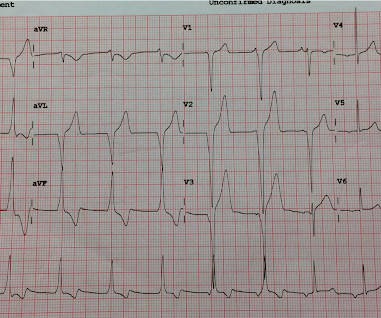
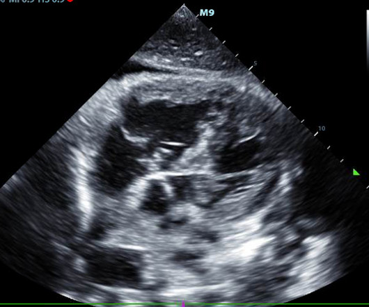
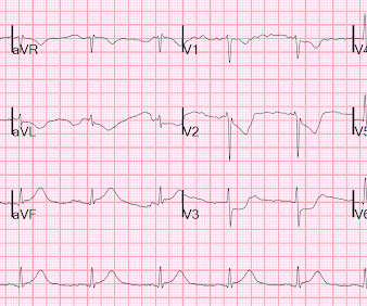
Bedside cardiac ultrasound showed moderately decreased LV function. She had an ECG recorded: This is left bundle branch block (LBBB), with appropriate proportional discordance. In the middle of the night, a "code" was called, and multiple rhythms like this were recorded. She was intubated. Modified QT = 440 - 65 = 375 ms.

EKG/ECG 115
article thumbnail

Four patients with chest pain and ‘normal’ ECG: can you trust the computer interpretation?

Dr. Smith's ECG Blog

All initial ECGs were labeled ‘normal’ or ‘otherwise normal’ by the computer interpretation, and below are the ECGs with the final cardiology interpretation. 1-3] But these studies were very short duration and used cardiology interpretation of ECGs or emergent angiography rather than patient outcomes.

EKG/ECG 118
Insiders

Sign Up for our Newsletter

This site is protected by reCAPTCHA and the Google Privacy Policy and Terms of Service apply.

Trending Sources

article thumbnail

Dynamic OMI ECG. Negative trops and negative angiogram does not rule out coronary ischemia or ACS.

Dr. Smith's ECG Blog

Here is his ED ECG at triage: Obvious high lateral OMI that does not quite meet STEMI criteria. He had a previous ECG on file: Proving the findings are new The cath lab was activated. Bedside cardiac ultrasound with no obvious wall motion abnormalities. Bedside cardiac ultrasound with no obvious wall motion abnormalities.

EKG/ECG 121
article thumbnail

emDOCs Podcast – Episode 98: Post ROSC Mental Model

EMDocs

Check the pulse RSI= Resuscitation Sequence Intubation Hypoxia, Hypotension, and Acidosis are the reason patients code during/post intubation These patients are super high risk for all 4 Optimize first pass success – Induction agent + paralytic Unconscious patients will still have muscle tone Induction Ketamine or Etomidate at half doses (i.e.,

article thumbnail

Three normal high sensitivity troponins over 4 hours with a "normal ECG"

Dr. Smith's ECG Blog

The documentation does not describe any additional details of the history. The following ECG was obtained. ECG 1 What do you think? The ECG shows sinus bradycardia but is otherwise normal. There is TWI in lead III, but this can be seen in normal ECGs. The following ECG was obtained around midnight.

EKG/ECG 119
article thumbnail

Putting Clinical Gestalt to Work in the Emergency Department

ACEP Now

Our nurse did not study Paul Ekman’s Facial Action Coding System for Action Units to code “fear” in the patient’s face. Does that normal troponin and ECG obviate the need for cardiology consultation for my patient with a concerning story for acute coronary syndrome? A testicular ultrasound confirmed restored blood flow.

article thumbnail

Dr. Elsburgh Clarke Was Among First to Specialize in Emergency Medicine

ACEP Now

A closer look, though, also shows the technology of the daya bulky, two-way radio for communicating with EMS, metal gurneys, glass saline bottles, and portable ECG monitors the size of a small shopping cart. Notice the use of the medical anti-shock trousers and the ECG machine. Now, we have ultrasound or CT scans to confirm.