Remove Documentation/Coding Remove EKG/ECG Remove Sepsis
article thumbnail

Torsade in a patient with left bundle branch block: is there a long QT? (And: Left Bundle Pacing).

Dr. Smith's ECG Blog

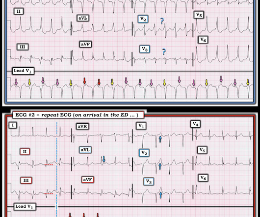
She was managed for sepsis with antibiotics including azithromycin, had hypotension with arterial and central lines placed and pressors. She had an ECG recorded: This is left bundle branch block (LBBB), with appropriate proportional discordance. Bedside cardiac ultrasound showed moderately decreased LV function.

EKG/ECG 117
article thumbnail

ECG Blog #366 — Diltiazem didn't work.

Ken Grauer, MD

The ECG and long lead II rhythm strip in Figure-1 — was obtained from a COVID positive patient with persistent tachycardia not responding to Diltiazem. Figure-1: The initial ECG — obtained from a patient with persistent tachycardia. ( To improve visualization — I've digitized the original ECG using PMcardio ).

EKG/ECG 195
Insiders

Sign Up for our Newsletter

This site is protected by reCAPTCHA and the Google Privacy Policy and Terms of Service apply.

article thumbnail

emDOCs Podcast – Episode 98: Post ROSC Mental Model

EMDocs

Check the pulse RSI= Resuscitation Sequence Intubation Hypoxia, Hypotension, and Acidosis are the reason patients code during/post intubation These patients are super high risk for all 4 Optimize first pass success – Induction agent + paralytic Unconscious patients will still have muscle tone Induction Ketamine or Etomidate at half doses (i.e.,

article thumbnail

Putting Clinical Gestalt to Work in the Emergency Department

ACEP Now

Our nurse did not study Paul Ekman’s Facial Action Coding System for Action Units to code “fear” in the patient’s face. Does that normal troponin and ECG obviate the need for cardiology consultation for my patient with a concerning story for acute coronary syndrome? We should respect our own and others’ gestalt assessments.

article thumbnail

What are treatment options for this rhythm, when all else fails?

Dr. Smith's ECG Blog

The below ECG was recorded. The ECG shows obvious STEMI(+) OMI due to probable proximal LAD occlusion. This ECG does not have the typical ST-vector of an LAD occlusion. See below for Ken Grauer Comment on the initial ECG: == On arrival, another ECG was recorded: There appears to have been quite a bit of spontaneous reperfusion!

EKG/ECG 132
article thumbnail

Medical Malpractice Insights: A Rare Presentation – Groin pain? Nec fasc? Diabetes? Appendicitis?

EMDocs

Exam is normal except for tenderness as documented in the diagram. EKG shows atrial fibrillation with a rate of 169. Sepsis is diagnosed and antibiotics started for the first time. High level amputation is considered, but she dies of sepsis and multiorgan failure on hospital day 7. I just failed to document it.

article thumbnail

Proning for ARDS

Northwestern EM Blog

There are several etiologies including viral pneumonia, bacterial pneumonia, sepsis, trauma, and pancreatitis. Remove ECG leads and patches. Reattach EKG leads to back. Document thorough skin assessment every nursing shift, and inspect weight-bearing ventral surfaces. Leave most of the sheet hanging. Suction as needed.