Remove Documentation/Coding Remove EKG/ECG Remove Hyperthermia / Hypothermia
article thumbnail

An elderly male with acute altered mental status and huge ST Elevation

Dr. Smith's ECG Blog

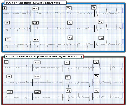
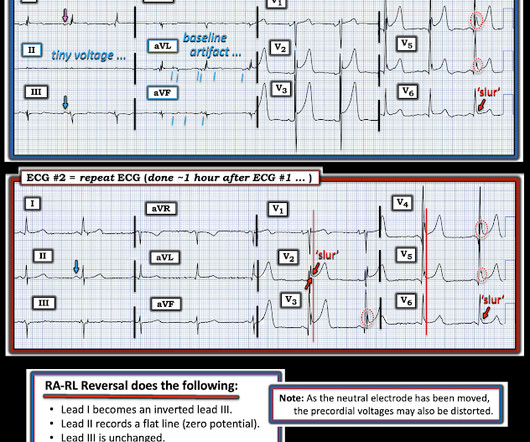
EKG on arrival to the ED is shown below: What do you think? The providers documented concern for ST elevation in the precordial and lateral leads as well as a concern for hyperkalemic T waves in the setting of succinylcholine administration. limb lead reversal is now resolved) Unfortunately, QOH V1 got tricked by this second ECG!

EKG/ECG 98
article thumbnail

Hyperthermia and ST Elevation

Dr. Smith's ECG Blog

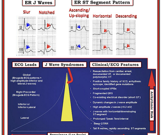
An initial EKG was obtained: Computer read: sinus tachycardia, early acute anterior infarct. Here is her prior EKG: When compared to the old EKG – Q waves present before, TWI in aVR present before, but all other changes are new. What is the differential for this EKG? T wave inversion III, aVR, TWF in aVF. Is this an OMI?

Insiders

Sign Up for our Newsletter

This site is protected by reCAPTCHA and the Google Privacy Policy and Terms of Service apply.

article thumbnail

SGEM#391: Is it Time for a Cool Change (Hypothermia After In-Hospital Cardiac Arrest)?

The Skeptics' Guide to EM

You are tidying your things […] The post SGEM#391: Is it Time for a Cool Change (Hypothermia After In-Hospital Cardiac Arrest)? You are tidying your things in anticipation of the arrival of the dayshift when a code blue is called. A post-arrest ECG doesn’t show any signs of STEMI. Are we supposed to be starting hypothermia?”

article thumbnail

ECG Pointers: Recurrent and Refractory Torsades de Pointes

EMDocs

An ECG is performed and is shown below: Figure 1. Adapted from Dr. Smith’s EKG Blog. Another ECG is obtained and shown below. Source: [link] As you are calling the ICU and cardiology team, the patient has recurrence of her symptoms and repeat ECG shows return of the PVT. She denies chest pain. What do you do?

EKG/ECG 71
article thumbnail

A man in his 70s with weakness and syncope

Dr. Smith's ECG Blog

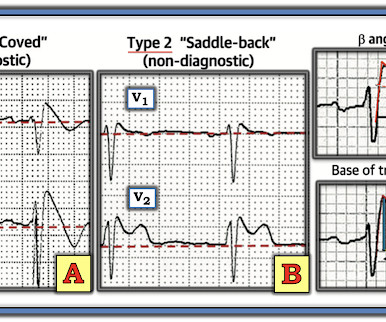
An ECG was performed in the ED at 1554: Original image unavailable, this is the only recorded scanned ECG available. In a patient with syncope and fever, this ECG looks more like Brugada. Smith comment: the ECG in question could be due to Brugada, even though there is a change from baseline. PM Cardio digitized version.

EKG/ECG 70
article thumbnail

Chemical Burns

Mind The Bleep

Circulation Assess heart rate, blood pressure, peripheral and central CRT, pulses and 3 lead ECG. Exposure Expose the patient in a systematic manner while keeping remaining body areas covered e.g. 1 limb at a time, to reduce the risk of hypothermia. Administer high-flow oxygen if indicated and consider arterial blood gas ( ABG ).

Burns 52
article thumbnail

Grand Rounds Recap 5.3.23

Taming the SRU

then need further evaluation Usually with CTA imaging If normal physical exam & ABI>0.9,