Remove Documentation/Coding Remove EKG/ECG Remove EMS
article thumbnail

Torsade in a patient with left bundle branch block: is there a long QT? (And: Left Bundle Pacing).

Dr. Smith's ECG Blog

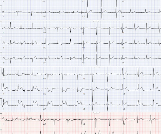
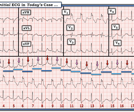
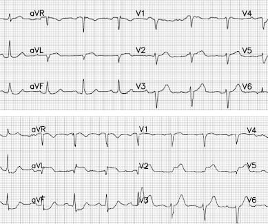
She had an ECG recorded: This is left bundle branch block (LBBB), with appropriate proportional discordance. Dodd KW, Elm KD, Dodd EM, Smith SW. In the middle of the night, a "code" was called, and multiple rhythms like this were recorded. CT of the chest showed no pulmonary embolism but bibasilar infiltrates. 2014;11:22732277.

EKG/ECG 116
article thumbnail

Another deadly triage ECG missed, and the waiting patient leaves before being seen. What is this nearly pathognomonic ECG?

Dr. Smith's ECG Blog

Pain improved to 1/10 after EMS administers 324 mg aspirin and the following EKG is obtained at triage. If this EKG were handed to you to screen from triage without any clinical information, what would you think? Do you appreciate any dynamic changes compared to the patient’s prior EKG? What do you think?

EKG/ECG 139
Insiders

Sign Up for our Newsletter

This site is protected by reCAPTCHA and the Google Privacy Policy and Terms of Service apply.

Trending Sources

article thumbnail

EM@3AM: Brugada Syndrome

EMDocs

We’ll keep it short, while you keep that EM brain sharp. Her presenting EKG is shown below. Clinical features Patients often present after an episode of sudden syncope, although Brugada syndrome can also be found on a routine EKG. ECG to evaluate for arrhythmia. Neurological exam is also unremarkable.

EMS 105
article thumbnail

Chest pain with serial ECGs – can you guess the sequence?

Dr. Smith's ECG Blog

Below are serial ECGs focusing on the inferior leads and aVL. First, what’s the interpretation of each ECG on its own? #1 2 Normal ECG #3. 2 Normal ECG #3. But 90 minutes later troponin returned at 70ng/L (normal <26 in males and <16 in females), and a repeat ECG was done (ECG#2) for recurring chest pain.

EKG/ECG 112
article thumbnail

Is OMI an ECG Diagnosis?

Dr. Smith's ECG Blog

What do you think of the ECG, and does it matter? I sent this to the Queen of Hearts So the ECG is both STEMI negative and has no subtle diagnostic signs of occlusion. 2] This is because, contrary to Bayesian reasoning, the STEMI paradigm is named after and defined by one part of one test: ST elevation on ECG. But only 6.4%

EKG/ECG 121
article thumbnail

The Expert Witness re-visits a chest pain Malpractice case using the Queen of Hearts

Dr. Smith's ECG Blog

Don't miss his analysis and assessment of the Queen of Hearts AI OMI ECG bot -- that assessment is at the very bottom of the post. Her first set of vitals were documented: BP 116/57 Pulse 94bpm Respiratory rate 24/min O2 sat 90% on room air Temp 97F She had been cleaning a Jeep in the sun, and was sunburned. But which artery?

EKG/ECG 92
article thumbnail

An elderly male with acute altered mental status and huge ST Elevation

Dr. Smith's ECG Blog

With EMS, patient had a GCS of 3 and was saturating 60% on room air. He improved to 100% with the addition of non-rebreather, however remained altered and was intubated by EMS with ketamine and succinylcholine. EKG on arrival to the ED is shown below: What do you think? or basilar ischemia. Potassium resulted as 4.9,

EKG/ECG 116