Remove Dislocations Remove Outcomes Remove Ultrasounds
article thumbnail

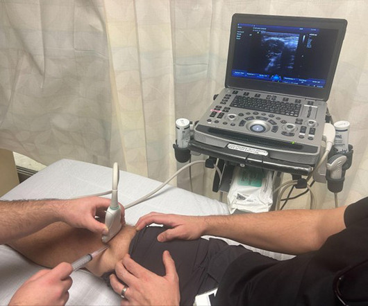
US Probe: Suprapatellar Recess Injection for Chronic Knee Pain in the Emergency Department – An Effective Approach for Relief

EMDocs

Author: Abdo Zeinoun MD, Clinical Ultrasound Fellow, Department of Emergency Medicine, Rutgers New Jersey Medical School // Reviewed by: Stephen Alerhand, MD ( @SAlerhand) ; Steve Fields, MD Patient Case A 60 year-old male with a history of hypertension presents with worsening right knee pain over the last 3 days. Why Use Ultrasound Guidance?

article thumbnail

emDOCs Podcast – Episode 118: Lisfranc Injury

EMDocs

Exist along a spectrum: minor subluxation to fracture and dislocation. Injuries range from sprain to fracture-dislocation; divided into low energy and high energy. Fracture displacement, dislocation -> reduce if displaced/dislocated (hang the foot by the toes using finger traps and then reduce); place in short leg posterior splint.

Insiders

Sign Up for our Newsletter

This site is protected by reCAPTCHA and the Google Privacy Policy and Terms of Service apply.

Trending Sources

article thumbnail

Grand Rounds Recap 1.3.24

Taming the SRU

R4 Case Follow-up: SCAD WITH dr. Martella Spontaneous Coronary Artery Dissection (SCAD) is a diagnosis confirmed via imaging: Coronary Angiography, Optical Coherence Tomography, Intravascular Ultrasound Therefore, treatment in the ED is the same as atherosclerotic ACS: ASA, heparin gtt and possible statin.

EKG/ECG 93
article thumbnail

Top Blogs of 2021

Northwestern EM Blog

Take-Home Point: Bedside pericardiocentesis is a rare but important procedure that can be performed emergently using ultrasound or landmark guidance. Take-Home Point: Knee dislocations are time-sensitive injuries that require rapid diagnosis and treatment because of the high rate of vascular complications.

article thumbnail

Grand Rounds Recap 3.8.23

Taming the SRU

mepivacaine (1-3 h) 1% lidocaine +/- epi (2-3h) 0.25% bupivacaine (2-3 h) 0.25-0.5% mepivacaine (1-3 h) 1% lidocaine +/- epi (2-3h) 0.25% bupivacaine (2-3 h) 0.25-0.5% mg/kg IV Versed: 0.2 mg/kg IM, 0.2 mg/kg IN (may repeat to max of 0.4 mg/kg IN), 0.2

article thumbnail

Diagnostics and Therapeutics: Managing Pneumothorax

Taming the SRU

As these patients are typically already at a high risk for poor health outcomes, secondary spontaneous PTX is often more serious in presentation and management more complex [11]. Blunt trauma can cause rib fracture or dislocation that may injure the visceral pleura. The "lung point": an ultrasound sign specific to pneumothorax.

article thumbnail

emDOCs Podcast – Episode 80: Traumatic Arthrotomy

EMDocs

Functional outcomes Severe soft tissue injuries and damage to the bony structures => poor functional outcomes. Can Ultrasound Identify Traumatic Knee Arthrotomy in a Cadaveric Model? The role of computed tomography in the assessment of open periarticular fractures associated with deep knee wounds. J Orthop Trauma.