This site uses cookies to improve your experience. To help us insure we adhere to various privacy regulations, please select your country/region of residence. If you do not select a country, we will assume you are from the United States. Select your Cookie Settings or view our Privacy Policy and Terms of Use.
Cookie Settings
Cookies and similar technologies are used on this website for proper function of the website, for tracking performance analytics and for marketing purposes. We and some of our third-party providers may use cookie data for various purposes. Please review the cookie settings below and choose your preference.
Used for the proper function of the website
Used for monitoring website traffic and interactions
Cookie Settings
Cookies and similar technologies are used on this website for proper function of the website, for tracking performance analytics and for marketing purposes. We and some of our third-party providers may use cookie data for various purposes. Please review the cookie settings below and choose your preference.
Strictly Necessary: Used for the proper function of the website
Performance/Analytics: Used for monitoring website traffic and interactions
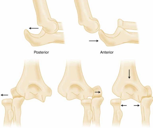
Elbow Dislocation Definition: Disarticulation of the proximal radius & ulna bones from the humerus Epidemiology: Incidence Second most common joint dislocation (after shoulder) in adults Most commonly dislocated joint in children Accounts for 10-25% of all injuries to the elbow ( Cohen 1998 ) Posterolateral is the most common type of dislocation (..)
Exist along a spectrum: minor subluxation to fracture and dislocation. of all fractures with an annual incidence of 1 per 55,000 persons. Injuries range from sprain to fracture-dislocation; divided into low energy and high energy. Big picture: look for bony widening, misalignment and fracture or avulsion.
Let’s talk about mandible dislocations & how we can reduce them. First, we need to take a look at the mandible anatomy – can refer back to this as we discuss mandible dislocation: So how does the mandible dislocate (also called temporomandibular joint dislocation)? Iatrogenic : ex.
Evaluating the limping child , though, requires us to ponder not only the common (ex, Toddler’s Fracture ), but also to be vigilant for the severe (ex, Septic Arthritis ). Mechanism of dislocation : [ Horan, 2006 ] Typically, the injury occurs when: The Knee is in Flexion and The Foot is rotated and plantar flexed.
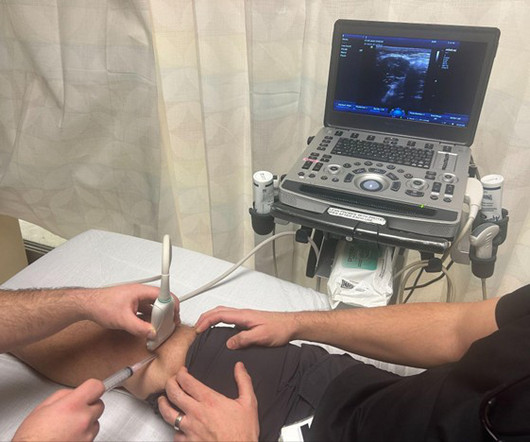
Diagnostic accuracy of point-of-care ultrasound (PoCUS) for shoulder dislocations and reductions in the emergency department: a diagnostic randomised control trial (RCT). BACKGROUND Shoulder injury and dislocations are common reasons for patients to present to the emergency department (ED) for evaluation. 39, 655–661 (2022).
Many vertebral fractures can be treated non-operatively. So I would like to concentrate on some papers that examined the use of back braces on patients who underwent pedicle screw fixation of their thoracic and/or lumbar spine fractures. Is postoperative bracing after pedicle screw fixation of spine fractures necessary?
X-ray shows mild joint space narrowing and osteophytes, with no fractures or dislocations. The contralateral knee is non-tender with full range of motion. He is neurovascularly intact distally. You reassess the patient, informing him of the negative x-ray findings, and his pain remains unchanged.
The post Ep 179 Hand Injuries – Finger Tip Injuries, Jersey Finger, PIP Dislocations, Metacarpal Fractures, Thumb Injuries, Tendon Lacerations appeared first on Emergency Medicine Cases.
Illustration by Yvonne Chow The Lisfranc Injury The lisfranc injury is any disruption of the joint and is a spectrum including ligamentous injury, dislocations and fractures. High energy mechanisms, such as MVCs or falls, are more likely to cause fracture-dislocations. Normal Lisfranc joint and ligament.
Most commonly caused by fracture or dislocation of vertebrae. Pathophysiology Primary injury happens at the time of the traumatic event or shortly after in the high cervical to mid-thoracic spine. This leads to descending sympathetic tracts being disrupted. Secondary spinal cord injury occurs hours to days after the initial insult.
Loss of cervical lordosis Concerning mechanism Remember: CT will help identify bony injury, however MRI is the better test to assess for ligamentous injury as well as spinal cord injury Below is an example of a Hangman’s Fracture (See Cervical Spine Fractures ). Moral of the Morsel You eyes may deceive you!
Triquetrum fracture / dislocation or carpal ligamentous injury stage III – stage II injury + dislocated triquetrum; can be associated … Continue reading →
Sternoclavicular (SC) joint dislocation SC joint dislocation can occur with anterior or posterior displacement of the medial clavicular head. Anterior dislocations are mostly caused by medial impact to the lateral shoulder. Anterior dislocations are more common and generally regarded as less serious.
On point-of-care ocular ultrasound of the affected eye using the linear probe a round, mobile structure was noted in the posterior chamber with an absence of the lens in its typical position, consistent with posterior lens dislocation. In this case, ophthalmology was consulted for lens dislocation. and a sensitivity of 96.8% (2).
Whether you’re interested in orthopaedics or not, knowledge of basic fracture management can be useful in any ED. Examination of a fractured limb Most patients will be in a lot of pain. If the limb is pale and pulseless this needs urgent referral to vascular as well as orthopaedics and the fracture needs reducing immediately.
Which proximal humerus fractures are likely to require surgical management? What is the best x-ray view to diagnose a sternoclavicular dislocation? What are the surgical indications for clavicle fractures? What are the surgical indications for clavicle fractures? and many more.
Join hosts Jeff Nusbaum, MD, and Nachi Gupta, MD on this episode of EMplify as they take you through the June 2018 issue of Emergency Medicine Practice: Managing Shoulder Injuries in the Emergency Department Fracture, Dislocation, and Overuse. Management of prehospital shoulder dislocation: feasibility and need of reduction.
Background Hip fractures are a very frequent presentation, even in non trauma centers. Adequate pain control, and early surgical treatment and mobilization are the main goals of hip fracture treatment; to reduce complications including infections, DVT and delirium.
Background: Many clinicians have transitioned from procedural sedation and analgesia (PSA) in favor of intra-articular lidocaine (IAL) to manage anterior shoulder dislocation. PMID: 36181665 Clinical Question: In patients with acute anterior shoulder dislocations, how does IAL compare to PSA for closed reduction?
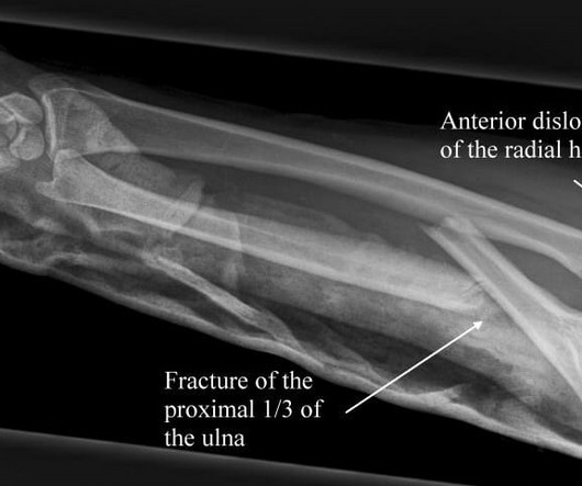
Monteggia fracture-dislocation. There is fracture involving proximal third of ulna & disruption of radio capitellar line indicating dislocation of the … Continue reading →
Eponym: Monteggia fracture (1812) ulna fracture, radial head dislocation Mike Cadogan Giovanni Battista Monteggia Giovanni Battista Montéggia (1762-1815) was an Italian surgeon.
The wrist x-rays show multiple fractures but the main injury is trans scaphoid perilunate dislocation. There are also fractures involving … Continue reading →
Pramod Chandru Network Five: Orthopaedics Network Five Emergency Medicine Journal Club Episode 22 - Orthopaedics reviewing papers on vascular injuries from knee dislocations, distal radius fractures and all things pelvic binders!
3,4 Risk factors include engaging in high energy activities, consumption of large amounts of alcohol, osteoporosis, and previous thoracic or lumbar fractures. 10 Spinal Fracture Classification 11 There are multiple different classification systems available. Spine fracture. The spinal canal houses the spinal cord. J Emerg Med.
It's all in the hips- fractures and dislocations. It's all in the hips- fractures and dislocations. Check out our interactive question bank podcast- the FIRST of its kind here: emrapidbombs.supercast.com. As Shakira says, the hips don't lie. So we've packed a lot of truthful information into this podcast. Episode 164.
It's all in the hips- fractures and dislocations. It's all in the hips- fractures and dislocations. Check out our interactive question bank podcast- the FIRST of its kind here: emrapidbombs.supercast.com. As Shakira says, the hips don't lie. So we've packed a lot of truthful information into this podcast. Episode 164.
Study Design A prospective study from a single ED in Belgium enrolled adults presenting to the ED within eight days after an acute injury with negative X-ray findings and a positive lever sign, Lachman test, or anterior drawer test, thus indicating clinical suspicion of ACL injury.
The patient reported a history of falls resulting in shoulder, rib, and left hip fractures in the past. Occult femur fracture Occult fractures are defined as fractures that cannot be detected by standard radiographic examination until weeks after the injury either due to lack of displacement or limitations of the imaging study.
3 Tenderness over the distal radial metaphysis after wrist injury is strongly suggestive of a distal radius fracture despite normal plain radiographs and fluoroscopic images. Children and older adults have weaker long bones than young adults and are more likely to sustain a distal radius fracture after a FOOSH than a carpal bone injury.
His x-ray imaging is seen below: View fullsize View fullsize Click on images above to enlarge Diagnosis: Volar Lunate Dislocation Lunate dislocation is an uncommon traumatic wrist injury that is often a result of high energy trauma from loading of a dorsiflexed wrist along with forced ulnar deviation (i.e References: Sherman, S.
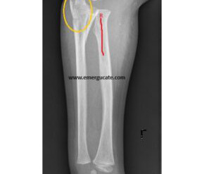
The elbow x-ray shows a fracture involving the olecranon process. There is also radial head dislocation (radiocapitellar line is broken). … Continue reading →
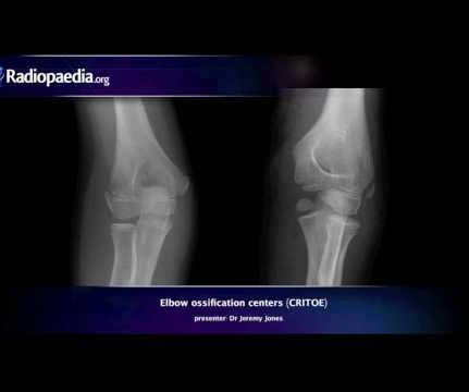
Pediatric Elbow Films: Putting It All Together Watch this dynamic video by Dr Jeremy Jone s from Radiopaedia : Fracture Saviors: Fat Pads and Drawn Lines These three things can save us: fat pads, the anterior humeral line, and the radiocapitellar line. If not, assume a fracture-dislocation.
The wrist x-rays show a trans-scaphoid peri-lunate dislocation. There are fractures involving the radial & ulnar styloid, mid-scaphoid, and triquetrum … Continue reading →
Final protocol will be published on Taming’s Clinical Guidelines in the coming months panorex WITH dr. urbanowicz Orthopantomography (OPG): specially protocoled panoramic radiographic tool which can visualize the teeth, mandible, upper limits of maxilla Convenient, inexpensive and rapid way to evaluate gross anatomy of jaws and related pathology Limitations: (..)
Monteggia fracturedislocation was first described in 1814 by Giovanni Battista Monteggia, 81 years before Roentgen described use of Xrays for medical … Continue reading →
Arun Sayal and Hossein Mehdian answer questions such as: When should we suspect a spontaneously reduced knee dislocation? Do all patients suspected of a spontaneous knee dislocation require a CT angiogram to rule out vascular injury? Which patients with a low energy mechanism are at risk for knee dislocation and vascular complications?
Beyde, artiga, and vaishnav Shoulder Dislocations Shoulder dislocations are easily diagnosed via ultrasound, with high sensitivity and specificity. Shoulder reductions can often be performed without sedation using an intra-articular injection.
Triquetrum chip fractures, scapholunate injuries, hook of the hamate fractures, and of course, scaphoid fractures can be easily missed with serious consequences for our patients.
In this main episode podcast we discuss the pitfalls in the diagnosis and management of elbow injuries and answer questions such as: What is an easy way to remember the surgical indications for radial head fractures? Why is it so important to assess for the extensor mechanism on physical exam for patients with olecranon fractures?
Maybe it's dislocated? It's a bit of a departure from what we've described above--he mainly goes right at a shaft fracture--but it's the same general idea: As always, these posts are for EDUCATIONAL PURPOSES ONLY. Ultrasound-Guided Three-In-One Nerve Block for Femur Fractures. Maybe he's broken his hip? doi:10.1111/acem.12154
We organize all of the trending information in your field so you don't have to. Join 5,000+ users and stay up to date on the latest articles your peers are reading.
You know about us, now we want to get to know you!
Let's personalize your content
Let's get even more personalized
We recognize your account from another site in our network, please click 'Send Email' below to continue with verifying your account and setting a password.


Let's personalize your content