This site uses cookies to improve your experience. To help us insure we adhere to various privacy regulations, please select your country/region of residence. If you do not select a country, we will assume you are from the United States. Select your Cookie Settings or view our Privacy Policy and Terms of Use.
Cookie Settings
Cookies and similar technologies are used on this website for proper function of the website, for tracking performance analytics and for marketing purposes. We and some of our third-party providers may use cookie data for various purposes. Please review the cookie settings below and choose your preference.
Used for the proper function of the website
Used for monitoring website traffic and interactions
Cookie Settings
Cookies and similar technologies are used on this website for proper function of the website, for tracking performance analytics and for marketing purposes. We and some of our third-party providers may use cookie data for various purposes. Please review the cookie settings below and choose your preference.
Strictly Necessary: Used for the proper function of the website
Performance/Analytics: Used for monitoring website traffic and interactions
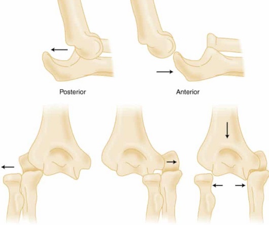
Elbow Dislocation Definition: Disarticulation of the proximal radius & ulna bones from the humerus Epidemiology: Incidence Second most common joint dislocation (after shoulder) in adults Most commonly dislocated joint in children Accounts for 10-25% of all injuries to the elbow ( Cohen 1998 ) Posterolateral is the most common type of dislocation (..)
Evaluating the limping child , though, requires us to ponder not only the common (ex, Toddler’s Fracture ), but also to be vigilant for the severe (ex, Septic Arthritis ). Mechanism of dislocation : [ Horan, 2006 ] Typically, the injury occurs when: The Knee is in Flexion and The Foot is rotated and plantar flexed.
To celebrate the end of trauma season ( is it ever really over? ), we here at the Ped EM Morsels Bakery have cooked up a morsel to remind you that pediatric trauma can be even more difficult than you think. Most commonly caused by fracture or dislocation of vertebrae. Never fear. Updated 2022 Mar 3]. In: StatPearls [Internet].
We’ll keep it short, while you keep that EM brain sharp. 3,4 Risk factors include engaging in high energy activities, consumption of large amounts of alcohol, osteoporosis, and previous thoracic or lumbar fractures. 10 Spinal Fracture Classification 11 There are multiple different classification systems available.
Loss of cervical lordosis Concerning mechanism Remember: CT will help identify bony injury, however MRI is the better test to assess for ligamentous injury as well as spinal cord injury Below is an example of a Hangman’s Fracture (See Cervical Spine Fractures ). Moral of the Morsel You eyes may deceive you! J Clin Orthop Trauma.
The post Ep 179 Hand Injuries – Finger Tip Injuries, Jersey Finger, PIP Dislocations, Metacarpal Fractures, Thumb Injuries, Tendon Lacerations appeared first on Emergency Medicine Cases.
A 10-year-old male with no past medical history presents to the Emergency Department (ED) by EMS for evaluation of an injury sustained while playing tackle football. Sternoclavicular (SC) joint dislocation SC joint dislocation can occur with anterior or posterior displacement of the medial clavicular head.
Which proximal humerus fractures are likely to require surgical management? What is the best x-ray view to diagnose a sternoclavicular dislocation? What are the surgical indications for clavicle fractures? What are the surgical indications for clavicle fractures? and many more.
Background: Many clinicians have transitioned from procedural sedation and analgesia (PSA) in favor of intra-articular lidocaine (IAL) to manage anterior shoulder dislocation. PMID: 36181665 Clinical Question: In patients with acute anterior shoulder dislocations, how does IAL compare to PSA for closed reduction?
Monteggia fracture-dislocation. There is fracture involving proximal third of ulna & disruption of radio capitellar line indicating dislocation of the … Continue reading →
The wrist x-rays show multiple fractures but the main injury is trans scaphoid perilunate dislocation. There are also fractures involving … Continue reading →
3 Tenderness over the distal radial metaphysis after wrist injury is strongly suggestive of a distal radius fracture despite normal plain radiographs and fluoroscopic images. Children and older adults have weaker long bones than young adults and are more likely to sustain a distal radius fracture after a FOOSH than a carpal bone injury.
Case by Sean Dyer PGY-3 EM Resident; Peer Reviewed by Dr. Christopher Hogrefe, Assistant Professor in Emergency Medicine at Northwestern University Feinberg School of Medicine The Case A 32 y/o male presents with wrist pain after being involved in a motor vehicle collision this afternoon. References: Sherman, S.
The elbow x-ray shows a fracture involving the olecranon process. There is also radial head dislocation (radiocapitellar line is broken). … Continue reading →
The wrist x-rays show a trans-scaphoid peri-lunate dislocation. There are fractures involving the radial & ulnar styloid, mid-scaphoid, and triquetrum … Continue reading →
Arun Sayal and Hossein Mehdian answer questions such as: When should we suspect a spontaneously reduced knee dislocation? Do all patients suspected of a spontaneous knee dislocation require a CT angiogram to rule out vascular injury? Which patients with a low energy mechanism are at risk for knee dislocation and vascular complications?
Monteggia fracturedislocation was first described in 1814 by Giovanni Battista Monteggia, 81 years before Roentgen described use of Xrays for medical … Continue reading →
EMS appears with a patient who just wiped out big time on the ice. Maybe it's dislocated? It's a bit of a departure from what we've described above--he mainly goes right at a shaft fracture--but it's the same general idea: As always, these posts are for EDUCATIONAL PURPOSES ONLY. Maybe he's broken his hip? doi:10.1111/acem.12154
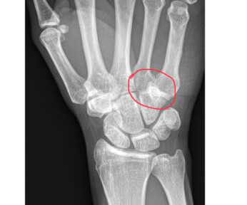
Triquetrum chip fractures, scapholunate injuries, hook of the hamate fractures, and of course, scaphoid fractures can be easily missed with serious consequences for our patients.
Furthermore, there is good evidence in the literature to support the use of ultrasound to accurately identify multiple shoulder pathologies including shoulder dislocation, rotator cuff tear, clavicle fracture and AC joint separation. The infraspinatus tendon will run just superior to these structures.
In this main episode podcast we discuss the pitfalls in the diagnosis and management of elbow injuries and answer questions such as: What is an easy way to remember the surgical indications for radial head fractures? Why is it so important to assess for the extensor mechanism on physical exam for patients with olecranon fractures?
We’ll keep it short, while you keep that EM brain sharp. Answer : Posterior dislocation of a periprosthetic hip Epidemiology: Total Hip Arthroplasty (THA) is the 4 th most common surgical procedures in the United States (2.3% 4 Dislocation is the most common complication after THA occurring at a rate of 0.2-10% 10% of patients.
There is an undisplaced olecranon fracture. The elbow x-ray shows haemathrosis as evidenced by elevated anterior & posterior fat pads. … Continue reading →
We’ll keep it short, while you keep that EM brain sharp. A 17-year-old right hand dominant male with no past medical history comes in complaining that he broke his right finger after being tackled at football practice. He has full ROM at the wrist, elbow, and shoulder. What is the diagnosis?
For instance, while pain resulting from extremity fractures and dislocations can often be relieved after reduction and splinting, abdominal pain poses a more elusive and complex scenario involving multiple contributing factors. Beyond the patient’s age and location of pain, we know very little of the cause or prognosis in this paper.
Location of the block will determine the morphology of the QRS (as a higher block may have a narrow QRS with a rate of 40-60bpm) Evaluation in the ED: basic labs including BMP and troponin, EKG, bedside echo, CXR Management: Atropine: push-dose 0.5-1mg,
A 44 year-old male with unknown past medical history came by emergency medical services (EMS) to the emergency department (ED) for an electrical injury and fall from a high voltage electrical pole. Per EMS, the patient was found at the bottom of a high voltage line with diffuse burns and amputation of his left forearm.
Jeff: With your initial stabilization underway, you can begin to gather a more thorough history either from bystanders or EMS if they are still present. Cerebral salt wasting syndrome, peripheral nerve lesions, spinal cord fracture, and cerebral hemorrhages have all been described.
How should pelvic fractures be identified in unstable trauma patients? What is the EM physician’s role in the stabilization of unstable pelvic injuries? Pelvic fractures can involve disruptions in any of the bony or ligamentous structures of the pelvic ring. While single-bone fractures can occur, they are far less common.
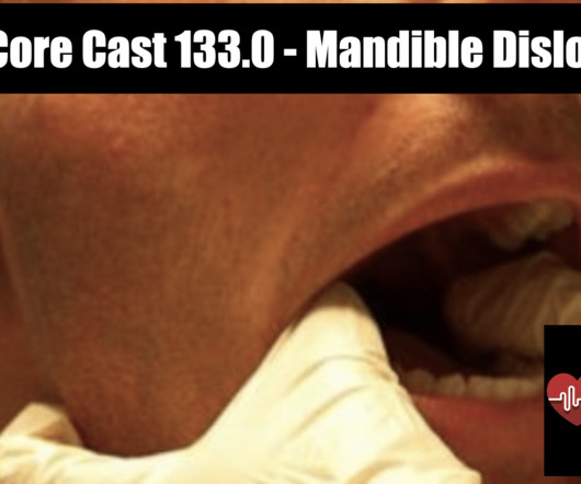
Temporomandibular (TMJ) Joint Dislocation Definition: Displacement of the mandibular condyle from the temporomandibular fossa. Epidemiology: Anterior dislocations are most common Mechanism Atraumatic (most common) from extreme mouth opening (yawning, eating, dental procedure, etc).
This means that we get so much hands-on experience doing procedures like fracture/dislocation reductions, complex laceration repairs, ENT procedures, and advanced eye exams. As EM residents, we support each other on shift and when signing out to ensure that folks are leaving to get out and enjoy their lives outside of work.
Ankle fractures are the third most common fracture in the ED [2] and more than 20,000 patients are seen in the ED for ankle sprains each day [3]. As an EM physician, it is important to have an understanding of the spectrum of ankle injuries and how these are appropriately evaluated. Range all joints.
As an EM physician, it is important to have an understanding of the spectrum of foot injuries and how these are appropriately evaluated. fractures of the talar body, talar neck, and calcaneus require high-impact trauma). Gross deformities often suggest fracture or dislocation. Range all joints.
Temporomandibular dislocation: a complication of tetanus. tetani infection is also indicated. Oral Surg Oral Med Oral Pathol Oral Radiol Endod. 2006;101(4):437-441. doi:10.1016/j.tripleo.2005.04.013 2005.04.013 Alfery DD, Rauscher LA. Tetanus: a review. Crit Care Med. 1979;7(4):176-181. J Trop Med Hyg. 1993;96(1):60-61.
We organize all of the trending information in your field so you don't have to. Join 5,000+ users and stay up to date on the latest articles your peers are reading.
You know about us, now we want to get to know you!
Let's personalize your content
Let's get even more personalized
We recognize your account from another site in our network, please click 'Send Email' below to continue with verifying your account and setting a password.


Let's personalize your content