This site uses cookies to improve your experience. To help us insure we adhere to various privacy regulations, please select your country/region of residence. If you do not select a country, we will assume you are from the United States. Select your Cookie Settings or view our Privacy Policy and Terms of Use.
Cookie Settings
Cookies and similar technologies are used on this website for proper function of the website, for tracking performance analytics and for marketing purposes. We and some of our third-party providers may use cookie data for various purposes. Please review the cookie settings below and choose your preference.
Used for the proper function of the website
Used for monitoring website traffic and interactions
Cookie Settings
Cookies and similar technologies are used on this website for proper function of the website, for tracking performance analytics and for marketing purposes. We and some of our third-party providers may use cookie data for various purposes. Please review the cookie settings below and choose your preference.
Strictly Necessary: Used for the proper function of the website
Performance/Analytics: Used for monitoring website traffic and interactions
The medics recorded an ECG: There is STE in V1-V3 and aVL, with reciprocal ST depression in II, III, aVF. An ECG was recorded: Now you can see what the medics could not: The QRS is enormous. ECG 3 hours later was unchanged He was not started on heparin as type II MI was favored over NonSTEMI as the etiology of his troponin elevation.
EKG shows atrial fibrillation with a rate of 169. Hospital Inpatient : R thigh is indurated and blistered but abdomen is all but ignored. Hospital Inpatient : R thigh is indurated and blistered but abdomen is all but ignored. On the 2nd hospital day, she becomes obtunded and is intubated. Glucose is 490, CO2 8, and pH 7.26
Other causes of sickling: acidosis, dehydration, inflammation, infection, fever, and blood stasis Sickling leads to vascular occlusion, end-organ ischemia, and decreased RBC lifespan, which, in turn, leads to pain crisis, acute anemia, sequestration, infection, and acute chest syndrome (ACS.) ECG: Evaluate for ischemia, right heart strain.
Other causes of sickling: acidosis, dehydration, inflammation, infection, fever, and blood stasis. ECG: Evaluate for ischemia, right heart strain. Fluid management Goal is euvolemia Dehydration – needs IV fluid resuscitation. HbS has reduced solubility in the setting of hypoxia, leading to sickling of the RBCs.
Her initial EKG is below. The following EKG was obtained after administration of adenosine. Unfortunately, shortly after this EKG was obtained, the patient returned to SVT. Same as initial ECG. ie, dehydration, periods of increased stress or anxiety, stimulants such as caffeine, alcohol, recreational drugs, etc.);
On arrival, here is his initial ED ECG, and is identical to the prehospital ECG and at the same rate: There is a regular, wide complex. The patient ' s baseline ECG looks exactly the same, except that it is slower (93) and the p-waves are obvious. Maybe the patient has dehydration, sepsis, hemorrhage, or PE.
They gave him water with salt, as he thought he was dehydrated." Vitals were obtained, and placed on cardiac monitor, including a 12 lead EKG." Transmitted to hospital with PCI." "ER There was no post cath ECG , which if unchanged would absolutely confirm that this is his baseline ECG. Pattern looked to be BER.
Note 2 other similar cases at the bottom that come from my book, The ECG in Acute MI. Case While I was busy seeing patients, a resident brought me this ECG of a 60-something with a history of syncope only. There was no chest pain or SOB at the tim of the ECG: Computerized QTc is 464 ms A previous ECG from 8 years prior was normal.
An immediate 12-lead EKG was obtained: There is ST elevation in leads aVR and V1, with marked ST depression in I, II, III, aVF, V3-V6. DISCUSSION: The 12-lead EKG EMS initially obtained for this patient showed severe ischemia, with profound "infero-lateral" ST depression and reciprocal ST elevation in lead aVR. What should be done?
Written by Pendell Meyers We received a call from an outside hospital asking to transfer a "traumatic post arrest" patient. We asked for the ECGs to be faxed over while they prepared to transfer him. Prior ECG on file: Sinus tachycardia, imperfect baseline, otherwise unremarkable. There is no ECG available from this time.
It is a very common presentation or comorbidity to see in community services and general hospital. environmental factors, pain, infection, dehydration, electrolyte disturbance, head injury, medication etc) Does the individual have capacity? The symptoms of BPSD are listed below. If there is no benefit this should be stopped.
Her name is Emily Groopman, and she’s a current resident at Children’s National Hospital. GI symptoms are the second most common, and they include vomiting, food intolerance, food aversion, GERD, refractory to normal antireflux measures, diarrhea, and dehydration.
A blind weight should be obtained post-void with the patient in a hospital gown only (to prevent hidden weights), and the patients back to the weight display. I recommend an order set that includes ECG, glucose, creatinine, liver enzymes, lipase, amylase, electrolytes including calcium, magnesium, and phosphate ketones, and urinalysis.
neurologic damage and thrombosis) 20,21 Resuscitation Lactated ringers > Normal saline due to reduced need for urinary alkalinization 22 Place foley and target urine output >300 mL/hr Electrolyte abnormalities Hyperkalemia: Obtain an ECG Stabilization: Calcium gluconate 2 g over 5-10 min. 1997;42(3):470-475; discussion 475-476.
Nachi: Sometimes… Jeff: This month’s issue was authored by Mollie Williams, who is the EM residency program director at the Brooklyn Hospital Center. Jeff: The hyperemetic phase lasts 24-48 hours and can lead to dehydration, electrolyte abnormalities, and weight loss. This is often associated with an inability to tolerate po.
Also consider non-hemorrhagic volume depletion, dehydration : orthostatic vitals may uncover this [see Mendu et al. (3)]. Abnormal ECG – looks for cardiac syncope. Abnormal Electrocardiogram (ECG): Defined (San Fran syncope rule) as any new changes when compared to the last ECG or presence of non-sinus rhythm.
Bhana, MD (EM Resident Physician, University of Massachusetts/UMass Chan Medical School); Clarence Kong, MD (Pain Fellow, Eastern Virginia Medical School – Virginia Health Sciences at Old Dominion University); Mani Hashemi, MD (EM Attending, HCA Florida Mercy Hospital); S.M. Epub 2021 Jan 2. PMID: 33098707. Fam AG, Smythe HA. J R Soc Med.
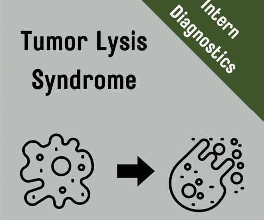
Initial work up when suspicious of this process should include ECG, chest x-ray, urinalysis, and the following labs: CBC, CMP, magnesium, phosphate, calcium, uric acid, VBG (for pH), and lactate dehydrogenase (10). Patients with TLS should be admitted to the hospital with concurrent management from the oncology and renal teams.
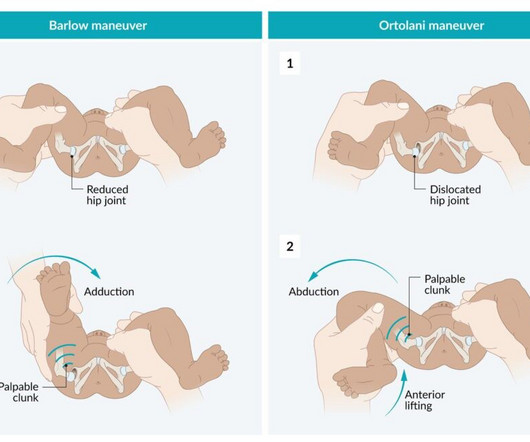
Every baby has a NIPE performed, either in hospital before discharge or at home by a community midwife. If the fontanelle is sunken then the baby may be dehydrated and if they are bulging then this can be a sign of birth injury or hydrocephalus. Request an ECG for the baby if ANY murmur is identified.
We organize all of the trending information in your field so you don't have to. Join 5,000+ users and stay up to date on the latest articles your peers are reading.
You know about us, now we want to get to know you!
Let's personalize your content
Let's get even more personalized
We recognize your account from another site in our network, please click 'Send Email' below to continue with verifying your account and setting a password.


Let's personalize your content