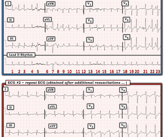
This site uses cookies to improve your experience. To help us insure we adhere to various privacy regulations, please select your country/region of residence. If you do not select a country, we will assume you are from the United States. Select your Cookie Settings or view our Privacy Policy and Terms of Use.
Cookie Settings
Cookies and similar technologies are used on this website for proper function of the website, for tracking performance analytics and for marketing purposes. We and some of our third-party providers may use cookie data for various purposes. Please review the cookie settings below and choose your preference.
Used for the proper function of the website
Used for monitoring website traffic and interactions
Cookie Settings
Cookies and similar technologies are used on this website for proper function of the website, for tracking performance analytics and for marketing purposes. We and some of our third-party providers may use cookie data for various purposes. Please review the cookie settings below and choose your preference.
Strictly Necessary: Used for the proper function of the website
Performance/Analytics: Used for monitoring website traffic and interactions
Check the pulse RSI= Resuscitation Sequence Intubation Hypoxia, Hypotension, and Acidosis are the reason patients code during/post intubation These patients are super high risk for all 4 Optimize first pass success – Induction agent + paralytic Unconscious patients will still have muscle tone Induction Ketamine or Etomidate at half doses (i.e.,
Hypothermic Arrest In general, hypothermic patients in cardiac arrest should be aggressively resuscitated. Patients can have excellent outcomes despite prolonged resuscitation. 2,3 If the patient meets criteria for resuscitation, they generally are not declared dead until their core temperature is above 32℃ (“warm and dead”).
What factors need to be taken into consideration to decide on when to terminate resuscitation of the cardiac arrest patient - when to call the code? The post Ep 170 Cardiac Arrest – PoCUS Integration, Communication Strategies, E-CPR, Calling the Code appeared first on Emergency Medicine Cases. and many more.
ultrasound grand rounds: bedside dvt studies - family presence in the ed/icu - r1 clinical knowledge: aicd - r3 small groups: difficult airway management Ultrasound grand rounds: DVT studies WITH Dr. minges Why should we perform bedside DVT studies in the ED? ETT onto a fiberoptic scope.
Unresponsive patients with undetectable MAP or EtCO2 less than 20 should undergo CPR. TEE can be helpful in guiding resuscitation if available. Factors that improve survival rates include cardiac activity on ultrasound, initial shockable rhythm, witnessed arrest, extremity only trauma, and bystander CPR.
Resuscitation , Volume 181,2022,Pages 140-146,ISSN 0300-9572, [link] Scott Weingart, MD FCCM. Available from: [link] Siamak Moayedi, Priya Patel, Nicholas Brady, Michael Witting, Timm-Michael L. Anteroposterior pacer pad position is better than anterolateral for transcutaneous cardiac pacing.” EMCrit 310 – Transvenous Pacemakers. EMCrit Blog.
Arrival at time 0 ECG 7 min Roomed in hallway at 17 min Moved to room with monitor at 37 min The patient was seen briefly by the physician, who then went to get an ultrasound machine. Resuscitative attempts were initiated quickly. VF was refractory to amiodarone, lidocaine, double-sequential defibrillation, esmolol, etc.
It might be better to consider traumatic cardiac arrest as a completely different disease eg LOST: Low Output State due to Trauma The 2015 European Resuscitation Council and UK Resuscitation Council Algorithms for Traumatic Cardiac Arrest: To read the whole ERC guideline on special circumstances cardiac arrest including trauma, click here.
ECMO Flow was achieved after approximately 1 hour of high quality CPR. The ways to tell for certain include intravascular ultrasound (to look for extra-luminal plaque with rupture) or "optical coherence tomography," something I am entirely unfamiliar with. After good ECMO flow was established, she was successfully defibrillated.
His primary interests are resuscitation, prehospital critical […] The post SGEM#231: You’re So Vein – IO vs. IV Access for OHCA first appeared on The Skeptics Guide to Emergency Medicine. His primary interests are resuscitation, prehospital critical care, airway management, and point-of-care ultrasound.
He underwent immediate CPR, was found to be in ventricular fibrillation, and was successfully resuscitated. I do not have the post-resuscitation ECG. An emergency cardiac ultrasound could be very useful. The physician recorded this ECG, interpreted it as normal, and sent the patient home on an antacid.
EMplify November 2020 An Evidence-Based Approach to Nonoperative Management of Traumatic Hemorrhagic Shock in the Emergency Department Authors : Christopher Pitotti, MD, FACEP Jason David, MD Topics : The Lethal Triad Prehospital Care Tourniquets - Placement and Pitfalls Stop The Bleed Hemostatic Dressings TXA Temperature Management ED Assessment Shock (..)
CPR was initiated immediately. Decision was made to push tPA after approximately 25 minutes of CPR, and after approximately 25 minutes after tPA was given, O2 saturation increased to 97%, and the patient was no longer cyanotic, converted to normal sinus rhythm with anterior lateral T wave inversions with ST depressions." These include.
Common causes include central venous catheterization (subclavian or internal jugular), lung biopsy, barotrauma from PPV, thoracentesis, bronchoscopy, pacemaker insertion, CPR, and intercostal nerve block [12, 14]. Unsuspected tension pneumothorax as a hidden cause of unsuccessful resuscitation. Resuscitation. Ann Emerg Med.
A bedside ultrasound was done by the emergency physician, using Speckle Tracking. The first time I looked at this, from afar, I did not see it, but after drawing the lines, one can clearly see abnormal ST elevation very suggestive of high lateral STEMI. Unfortunately, that video is unavailable.
On arrival, the patient was in shock, was intubated, and had an immediate cardiac ultrasound. What does a heart look like on ultrasound when the EKG looks like that? Here you go: It's not the world's greatest cardiac ultrasound video, but it does appear to show poor function and low volume. They transported to the ED.
Bedside ultrasound showed no effusion and moderately decreased LV function, with B-lines of pulmonary edema. After resuscitation, he was found to have a 90% thrombotic lesion in the same saphenous vein graft to the right posterior descending artery. He was managed medically with Clopidogrel. He appeared to be in shock. mg/dL [1.03 0.16
Data that do not establish neurological risk stratification in the first 6 hours after CA include the patient’s age, duration of CPR, seizure activity, serum lactate level or pH, Glasgow motor subscore in patients who received NMB or sedation, pupillary function in patients who received atropine, and optic nerve sheath diameter (95.3%, 20/21).
Thankfully, that gentleman was successfully resuscitated despite no bystander CPR, and if you listen carefully, we hope to arm you with the tools to do so similarly. These aren’t your standard arrest patients though, they typically have many fewer comorbidities – so CPR tends to be more successful. Let’s move on to treatment.
A bedside cardiac ultrasound was normal, with no effusion. He underwent CPR, and regained a pulse after epinephrine, with an organized narrow complex rhythm at 140, but still with severe shock. He had the following EKG recorded: Low voltage, suggests effusion. see Ken's discussion of low voltage below) There is a QS-wave in V2.
Nachi: And if the prehospital team is lucky enough, or maybe unlucky enough, i don’t know, to have a credentialed provider who can perform ultrasound for those suspected of having a blunt cardiac injury, the general prehospital data on ultrasound is sparse. Jeff: Great, let’s move onto ED care, beginning with the H&P.
In December 2014, ESEM brought together a group of local emergency physicians with specialized training in emergency ultrasound, establishing the emergency ultrasound committee, a key step in ESEM’s mission to maintain the highest practice standards and promote professional growth in emergency medicine within the UAE.
His main areas of interest are Critical Care, Ultrasound, Prehospital Resuscitation, and Medical Education. CPR was started immediately after the coaching staff could not find a pulse and EMS arrived to find the patient in cardiac arrest.
Article 1: Is lung ultrasound a viable alternative to chest x-ray in diagnosing community-acquired pneumonia? Diagnostic accuracy of point-of-care lung ultrasound for community-acquired pneumonia in children in ambulatory settings: A systematic review and meta-analysis. Ultrasound. 2024 Oct 29; Whats it about? Whats it about?
We organize all of the trending information in your field so you don't have to. Join 5,000+ users and stay up to date on the latest articles your peers are reading.
You know about us, now we want to get to know you!
Let's personalize your content
Let's get even more personalized
We recognize your account from another site in our network, please click 'Send Email' below to continue with verifying your account and setting a password.
Let's personalize your content