This site uses cookies to improve your experience. To help us insure we adhere to various privacy regulations, please select your country/region of residence. If you do not select a country, we will assume you are from the United States. Select your Cookie Settings or view our Privacy Policy and Terms of Use.
Cookie Settings
Cookies and similar technologies are used on this website for proper function of the website, for tracking performance analytics and for marketing purposes. We and some of our third-party providers may use cookie data for various purposes. Please review the cookie settings below and choose your preference.
Used for the proper function of the website
Used for monitoring website traffic and interactions
Cookie Settings
Cookies and similar technologies are used on this website for proper function of the website, for tracking performance analytics and for marketing purposes. We and some of our third-party providers may use cookie data for various purposes. Please review the cookie settings below and choose your preference.
Strictly Necessary: Used for the proper function of the website
Performance/Analytics: Used for monitoring website traffic and interactions
Ultrasound during cardiac arrest has quickly become standard. Initially, data suggested that the use of ultrasound during arrest increased pauses between compressions which worsens outcomes. Finally, patients with PEA and cardiac standstill on ultrasound have a 0.0%-0.6% Yours in ultrasounding, Shivam
in the paper but 2.7% to ≈0.99 (p<0.001) Mean MPI/Tei Index≈ 0.47 in the paper but 2.7% to ≈0.99 (p<0.001) Mean MPI/Tei Index≈ 0.47 in the paper but 2.7% to ≈0.99 (p<0.001) Mean MPI/Tei Index≈ 0.47 to 4.0mg/hr typically given in EKOS therapy (See Below). to 4.0mg/hr typically given in EKOS therapy (See Below).
Patients can have excellent outcomes despite prolonged resuscitation. VF/asystole), a pulse cannot be identified via Doppler ultrasound for a full minute, or if lack of organized cardiac activity is confirmed on bedside echocardiogram. This is a good time to utilize an arterial line or use the ultrasound to find a central pulse.
ultrasound grand rounds: bedside dvt studies - family presence in the ed/icu - r1 clinical knowledge: aicd - r3 small groups: difficult airway management Ultrasound grand rounds: DVT studies WITH Dr. minges Why should we perform bedside DVT studies in the ED? ETT onto a fiberoptic scope.
The conclusion from that episode was it’s still uncertain if using etomidate decreases the patient-oriented outcome of survival with good neurologic function in critically ill patients requiring emergent endotracheal intubation. The most recent discussion was about the use of etomidate as an induction agent ( SGEM#405 ).
There was no bystander CPR. Bedside ED ultrasound showed exceedingly poor global LV function, and no B lines. The patient awoke and had a good outcome! Medics found him in ventricular fibrillation. He was unidentified and there were no records available After 7 shocks, he was successfully defibrillated and brought to the ED.
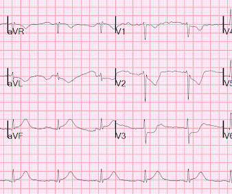
I sent it to 5 of my OMI friends without any clinical information or outcome and all 5 independently responded with exactly the same diagnosis: "reperfused inferior OMI". 3-vessel disease can make resuscitation very difficult, since CPR does not perfuse diseased vessels as well as one would like.
His primary interests are resuscitation, prehospital critical care, airway management, and point-of-care ultrasound. Bystander CPR is initiated prior to EMS arrival. However, no randomized trial has compared intravenous access to intraosseous access with a primary outcome of good neurologic function.
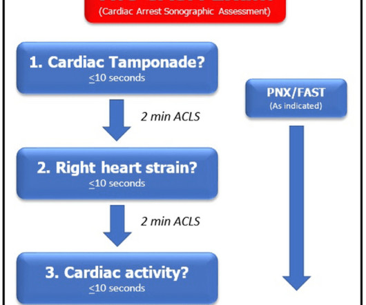
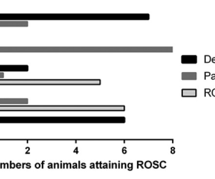
Ultrasound during TCA: Cureton et al. The heart of the matter: utility of ultrasound of cardiac activity during traumatic arrest. The outcomes from different resuscitative interventions in a haemorrhagic shock model in porcine model: From: Watts et al. Use of CPR in hemorrhagic shock, a dog model. 2012; 73: 102-10.
The paramedics achieve return of spontaneous circulation (ROSC) after CPR, advanced cardiac life support (ALCS), and Intubation. Her point-of-care ultrasound (POCUS) shows appropriate-appearing global ejection fraction and no marked wall motion abnormalities. The AHA has a statement with recommendations based on the available data.
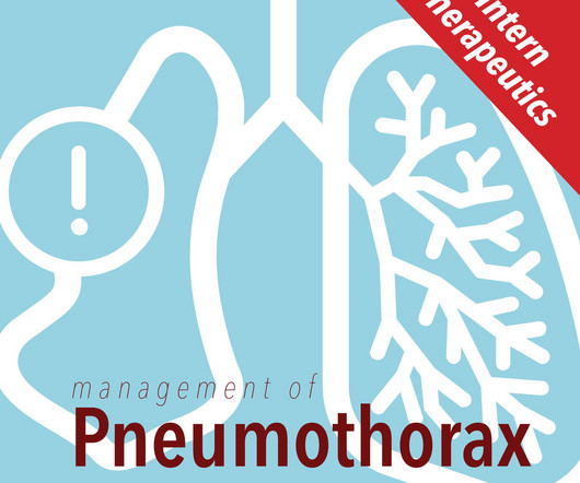
As these patients are typically already at a high risk for poor health outcomes, secondary spontaneous PTX is often more serious in presentation and management more complex [11]. Accuracy of Ultrasound in Diagnosis of Pneumothorax: A Comparison between Neonates and Adults-A Systematic Review and Meta-Analysis. Published 2020 Jul 23.
A bedside ultrasound was done by the emergency physician, using Speckle Tracking. It is highly associated with proximal LAD occlusion or severe left main ACS and with bad outcomes. Unfortunately, that video is unavailable. It appeared to show a lateral wall motion abnormality. So this is diagnostic of proximal LAD occlusion.
Data that do not establish neurological risk stratification in the first 6 hours after CA include the patient’s age, duration of CPR, seizure activity, serum lactate level or pH, Glasgow motor subscore in patients who received NMB or sedation, pupillary function in patients who received atropine, and optic nerve sheath diameter (95.3%, 20/21).
While Point of Care Ultrasound has made limited entry in prehospital care, largely with physician-led services and some Advanced Paramedics; it has largely been as a proof of concept rather than everyday care. These indices would have reinforced the clinical decision progress and avoided bad outcomes from unsuccessful estimates.
A bedside cardiac ultrasound was normal, with no effusion. He underwent CPR, and regained a pulse after epinephrine, with an organized narrow complex rhythm at 140, but still with severe shock. Cardiovascular Implications of Fatal Outcomes of Patients With Coronavirus Disease 2019 (COVID-19). 3–8 Shi et al. 2020; 10.
Nachi: And if the prehospital team is lucky enough, or maybe unlucky enough, i don’t know, to have a credentialed provider who can perform ultrasound for those suspected of having a blunt cardiac injury, the general prehospital data on ultrasound is sparse. Early defibrillation is linked to better outcomes. of those patients.
Bedside ultrasound showed no effusion and moderately decreased LV function, with B-lines of pulmonary edema. The patient stabilized and had a good outcome. Angio had shown some acute disease in the saphenous vein graft to the posterior descending artery off of the RCA. He was managed medically with Clopidogrel.
Article 1: Is lung ultrasound a viable alternative to chest x-ray in diagnosing community-acquired pneumonia? Diagnostic accuracy of point-of-care lung ultrasound for community-acquired pneumonia in children in ambulatory settings: A systematic review and meta-analysis. Ultrasound. 2024 Oct 29; Whats it about? Whats it about?
We organize all of the trending information in your field so you don't have to. Join 5,000+ users and stay up to date on the latest articles your peers are reading.
You know about us, now we want to get to know you!
Let's personalize your content
Let's get even more personalized
We recognize your account from another site in our network, please click 'Send Email' below to continue with verifying your account and setting a password.


Let's personalize your content