emDOCs Podcast – Episode 98: Post ROSC Mental Model
EMDocs
APRIL 2, 2024
Today on the emDOCs cast Brit Long interviews Zachary Aust on the use of a mental model in post ROSC patients. Episode 98: Post ROSC Mental Model What’s the problem?
This site uses cookies to improve your experience. To help us insure we adhere to various privacy regulations, please select your country/region of residence. If you do not select a country, we will assume you are from the United States. Select your Cookie Settings or view our Privacy Policy and Terms of Use.
Cookies and similar technologies are used on this website for proper function of the website, for tracking performance analytics and for marketing purposes. We and some of our third-party providers may use cookie data for various purposes. Please review the cookie settings below and choose your preference.
Used for the proper function of the website
Used for monitoring website traffic and interactions
Cookies and similar technologies are used on this website for proper function of the website, for tracking performance analytics and for marketing purposes. We and some of our third-party providers may use cookie data for various purposes. Please review the cookie settings below and choose your preference.
EMDocs
APRIL 2, 2024
Today on the emDOCs cast Brit Long interviews Zachary Aust on the use of a mental model in post ROSC patients. Episode 98: Post ROSC Mental Model What’s the problem?
Dr. Smith's ECG Blog
JULY 9, 2024
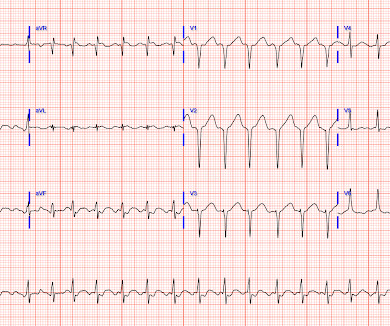
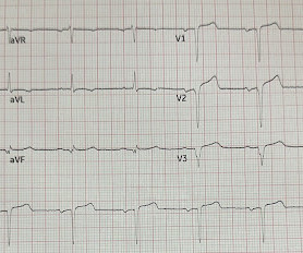
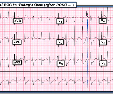
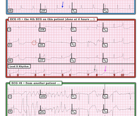
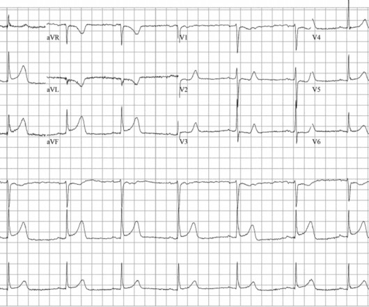
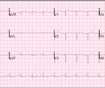
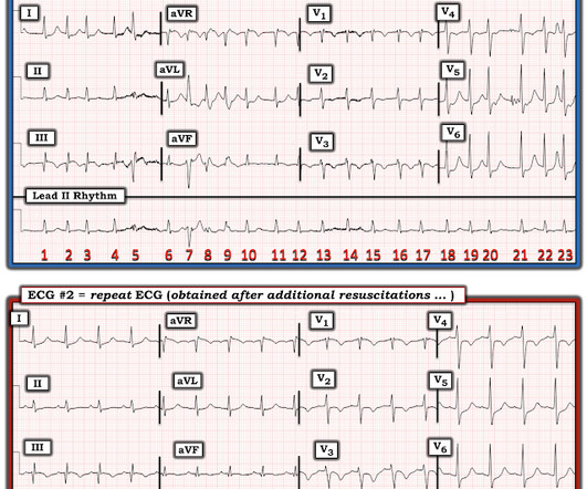
His family started CPR and called EMS, who arrived to find him in ventricular fibrillation. Here is his ECG after stabilization of vitals (at least 30 minutes since sustained ROSC). The ECG is diagnostic of acute LAD occlusion MI. Post angiogram ECG The patient was eventually able to be weaned off of ECMO and impella.
This site is protected by reCAPTCHA and the Google Privacy Policy and Terms of Service apply.
Dr. Smith's ECG Blog
JUNE 23, 2024
His daughter immediately started CPR and another family member called EMS. Here is his presenting ECG: ECG 1, t = 0 What do you think? His transfer packet included notes, labs, cath report, and ECG reports, but no actual ECG images. Smith's ECG Blog. He did not have access to ECG 1.
Dr. Smith's ECG Blog
SEPTEMBER 3, 2023
This ECG was recorded in triage. The computer interpretation is: “Sinus Brady with moderate intraventricular conduction delay, nonspecific t wave abnormality, abnormal EKG” What do you think? Case Continued The ECG findings were not recognized. Therefore, no matter the initial ECG, record serial ECGs.
Dr. Smith's ECG Blog
SEPTEMBER 11, 2024
Here is the case: Report from EMS was witnessed syncope, his son did CPR, but the patient had pulses when EMS arrived. Here is the ED ECG (a photo of the paper printout) What do you think? Instead — the "syncopal episode" prompted the patient's son to start CPR, and was associated with persistent hypotension.
Dr. Smith's ECG Blog
JULY 25, 2024
They started CPR. The patient was brought to the ED and had this ECG recorded: What do you think? The ECG shows severe ischemia, possibly posterior OMI. For clarity in Figure-1 — I've reproduced today's ECG — obtained following successful resuscitation of out-of-hospital cardiac arrest. He was defibrillated into VT.
RebelEM
SEPTEMBER 28, 2023
ECG Results: Repeat ECG 90min after tenecteplase indicated 70.3% ECG Results: Repeat ECG 90min after tenecteplase indicated 70.3% ECG Results: Repeat ECG 90min after tenecteplase indicated 70.3% The upper range of this time was 120 minutes from diagnostic ECG in the majority of patients.
Don't Forget the Bubbles
JULY 26, 2023
Add into this that the majority of children will be in normal sinus rhythm (NSR) by the time of assessment so to truly identify those who have something wrong we have to be confident in identifying arrhythmias where they are present and critical when analysing an ECG in NSR. All were examined and 98% had an ECG.
Dr. Smith's ECG Blog
APRIL 2, 2024
There was no bystander CPR. Here is the initial ED ECG. Other thought this was due to hyperkalemia, but the ECG does not have the appearance of hyperkalemia but does have the appearance of severe cardiomyopathy -- LBBB with very wide QRS) 3. ECG with LBBB and QRS of > 210 ms. An elderly man collapsed.
Don't Forget the Bubbles
JUNE 30, 2023
She was intubated at the scene and transported to your ED, with cardiopulmonary resuscitation (CPR) performed en route. You request a 12 lead ECG and repeat a blood gas, asking for it to be run on the PICU analyser. Your trusted nurse hands you the ECG: Paediatric ECG interpretation has never been your strong suit.
Emergency Medicine Cases
DECEMBER 8, 2020
In this month's EM Quick Hits podcast: Anand Swaminathan on lateral canthotomy, Emily Austin on pediatric cannabis poisoning, Reuben Strayer on an approach to hyperthermia, Brit Long on diagnosis and management of malignant otitis externa, Jesse McLaren on ECG diagnosis of occlusion MI in patients with BBB and Peter Brindley on prone CPR.
Dr. Smith's ECG Blog
APRIL 30, 2024
Bystander CPR. Here is the only prehospital 12-lead: Sinus tachycardia, somewhat wide QRS, Ischemia Here is the first ED ECG: What is the rhythm here? This ECG is pathognomonic of hyperkalemia , with wide QRS, very SHARPLY peaked T-waves, flat ST segments, RBBB pattern and large R-wave in aVR. Medics found patient in PEA arrest.
Dr. Smith's ECG Blog
JULY 14, 2023
Two recent interventions have proven in randomized trials to improve neurologic survival in cardiac arrest: 1) the combination of the ResQPod and the ResQPump (suction device for compression-decompression CPR -- Lancet 2011 ) and 2) Dual Sequential defibrillation. Finally, head-up CPR (which was not used here), makes for better resuscitation.
Dr. Smith's ECG Blog
OCTOBER 3, 2017
He received aspirin en route via EMS, and no EMS ECGs are available. Here is his ECG on presentation to the ED, approximately 45 minutes after onset of pain, but with pain improving since onset: What is your interpretation? I sent this ECG with no clinical information to Dr. Smith.
Dr. Smith's ECG Blog
MAY 19, 2020
ECMO Flow was achieved after approximately 1 hour of high quality CPR. A followup ECG was recorded 2 days later: No definite evidence of infarction. Smith — “ S hark F in” m orphology was noted on the initial monitoring strip, and initial 12-lead ECG. For clarity — I’ll again show the initial ECG done in the ED in Figure-1.
Taming the SRU
JUNE 26, 2024
Unresponsive patients with undetectable MAP or EtCO2 less than 20 should undergo CPR. Factors that improve survival rates include cardiac activity on ultrasound, initial shockable rhythm, witnessed arrest, extremity only trauma, and bystander CPR. One should ensure that the device is plugged in and an audible hum is heard.
Dr. Smith's ECG Blog
OCTOBER 10, 2022
CPR was initiated immediately. Initial ROSC was obtained, during which this ECG was obtained: What do you think? I sent it to 2 of my ECG nerd colleagues with no clinical information whatsoever, who instantly said: "Looks like afib with subendocardial ischemia and right heart strain pattern." "I
ACEP Now
JANUARY 4, 2025
A closer look, though, also shows the technology of the daya bulky, two-way radio for communicating with EMS, metal gurneys, glass saline bottles, and portable ECG monitors the size of a small shopping cart. Notice the use of the medical anti-shock trousers and the ECG machine. Click to enlarge.) Click to enlarge.)
RebelEM
JUNE 1, 2023
Bystander CPR, a known predictor of good outcomes, was more common in the SDCT cohort than in the standard care cohort. Control: Standard approach to diagnosis post-arrest but would be eligible for SDCT. There is no discussion of how many of the diagnoses made by SDCT could have been made clinically instead.
Dr. Smith's ECG Blog
APRIL 29, 2020
CPR was started immediately. I was texted this ECG in real time, but it turns out to actually be the 2nd one recorded in the ED. Just as interesting is EKG 1, 24 minutes before the first: What do you think here? Below, I post 4 more examples of ECGs that were recorded from patients who had Torsades. Is this inferior MI?
ACEP Now
DECEMBER 6, 2024
During initial assessment, an ECG was obtained and revealed ST-segment elevation (STE) in the inferior leads with ST depression anteriorly. Initial ECG demonstrating inferolateral ST segment elevation and anteroseptal depression, just prior to cardiac arrest. The ECG showed ST-segment elevation without obstructive coronary disease.
Dr. Smith's ECG Blog
FEBRUARY 8, 2022
On arrival, CPR was continued and core temperature was measured at 18° C (64.4° A 12-lead ECG was recorded: There is sinus rhythm with RBBB and right axis deviation. Guide to this image: 30 minutes later, at 31 C, this ECG was recorded: The RBBB has resolved. The Osborn waves on ECGs #1 and #2 are huge!
AED Leader
JULY 10, 2024
The Powerheart G5 is a user-friendly AED that provides real-time CPR feedback and can be used for adult and pediatric patients. It offers advanced features such as ECG monitoring and multiple rescue capabilities. It offers advanced features such as ECG monitoring and multiple rescue capabilities.
The Skeptics' Guide to EM
SEPTEMBER 11, 2021
The paramedics achieve return of spontaneous circulation (ROSC) after CPR, advanced cardiac life support (ALCS), and Intubation. The EKG shows sinus tachycardia with nonspecific changes and no ST segment elevations, Q waves, or hyperacute T waves. She has a history of hypertension and non-insulin dependent diabetes mellitus.
AED Leader
MAY 5, 2024
Let’s take a closer look at some of the critical features of Zoll AEDs: Zoll AED 3 : The Zoll AED 3 is a compact, user-friendly defibrillator that provides real-time CPR feedback and guidance. Zoll AED Plus : Zoll AED Plus is another popular model because of its unique CPR feedback technology.
Taming the SRU
OCTOBER 5, 2023
morbidity and mortality - quality improvement - research grand rounds - r1 clinical knowledge: pres/rcvs - r4 case follow-up: compartment syndrome Morbidity and Mortality WITH dr. finney Takotsubo Cardiomyopathy with COVID-19 Increasing incidence of Takotsubo Cardiomyopathy with the COVID-19 pandemic Morbidity and mortality is similar to that of ACS (..)
Dr. Smith's ECG Blog
FEBRUARY 2, 2017
Here is the ECG: Sinus tachycardia. This is an extremely dangerous ECG. When the ECG shows the effects of hypokalemia, it is particularly dangerous. "Administration of potassium may be empirical in emergent conditions. This patient presented with severe DKA. What do you think? What else? The K returned at 1.9
Thinking Critical Care
MAY 20, 2023
There’s going to be a buffet of workshops to pick and choose from that we are still putting together, from airway management to bedside procedures, ekg workshops, neuro exam workshop, wound dressing, and of course all kinds of POCUS. If you’re not personalizing your CPR, here’s the place to learn! This is gold.
Dr. Smith's ECG Blog
NOVEMBER 19, 2014
EMS recorded the following ECG: What do you see? In other words, the ST segment and T-wave should be discordant to (in the opposite direction of) the positive R' wave [ see the 2nd ECG of this post ]. A male in late middle age with a history of RCA stent 8 years prior complained of chest pain.
Dr. Smith's ECG Blog
AUGUST 9, 2019
Some total LAD occlusion manifest on the ECG similarly to some left main occlusions. Depending on where the STE vector of the LM occlusion shows up on the ECG, there may be either STE, or STD, or neither in aVR, and lead aVR will not be the important part of the ECG interpretation in these cases.
Dr. Smith's ECG Blog
OCTOBER 24, 2013
The physician recorded this ECG, interpreted it as normal, and sent the patient home on an antacid. He underwent immediate CPR, was found to be in ventricular fibrillation, and was successfully resuscitated. I do not have the post-resuscitation ECG. Did the ECG offer unseen hints? See explanation below. Lesson : 1.
Dr. Smith's ECG Blog
OCTOBER 28, 2013
It was witnessed, and CPR was performed by trained individuals. She arrived in the ED 37 minutes after 911 was called, with continuing CPR. The following 12-lead ECG was recorded at 11 minutes after ROSC. at the time of the ECG. at the time of the ECG. Here is the annotated ECG from above, with lines drawn.
EMDocs
JANUARY 8, 2024
For patients with OHCA, use of steroids during CPR is of uncertain benefit. Extracorporeal CPR Use of ECPR for patients with cardiac arrest refractory to standard ACLS is reasonable in select patients when provided within an appropriately trained and equipped system of care. COR 2b, LOE B-R. COR 2b, LOE C-LD.
EM SIM Cases
MAY 9, 2023
The team should begin CPR and follow the PALS pediatric bradycardia algorithm. After one round of CPR, the patient’s heart rate will increase and the consulting team should arrive. Normal paediatric ECG Despite intubation, the child remains hypoxemic and the team works through an approach to post-intubation hypoxemia.
Dr. Smith's ECG Blog
MARCH 9, 2017
This was her ECG (it is unclear if this was with or without pain): Computerized ECG Read: “Normal sinus rhythm. Normal ECG.” Overall this is a very quiet ECG. link] A repeat ECG was performed 1.5 hrs after the initial ECG) she develops severe, crushing chest pain. Don’t trust the computerized ECG reading.
Dr. Smith's ECG Blog
NOVEMBER 30, 2019
A prehospital ECG was recorded: Limb leads: Precordial Leads What is the therapy? What does a heart look like on ultrasound when the EKG looks like that? The followup ECG is here: Now the QRS is only slightly prolonged. A middle-aged woman was found down in her apartment unconscious. She was in shock with thready pulses.
AED Leader
JULY 19, 2023
Users are given audible instructions in English or Spanish that take them through the steps of CPR and shock delivery. This technology allows chest compressions to continue during an ECG, reducing pauses in CPR which may, in turn, improve survival rates. cprINSIGHT Analysis. cprINSIGHT Analysis. LIFELINKcentral.
Dr. Smith's ECG Blog
OCTOBER 20, 2013
Medics found her apneic and pulseless, began CPR, and she was found to be in asystole. Two prehospital 12-lead ECGs looked similar to this ED ECG: This shows diffuse ST depression (I, II, III, aVL, aVF, V3-V6) with reciprocal ST elevation in lead aVR. This ECG is diagnostic of diffuse subendocardial ischemia.
Don't Forget the Bubbles
OCTOBER 7, 2024
Suspected low voltage injuries (<1000v) should still be approached with an A-E assessment, but if 12 lead ECG and urinalysis are normal, it’s reasonable to discharge the patient. If the ECG is abnormal, the patient should be managed as if exposed to a high voltage (>1000v).
Dr. Smith's ECG Blog
JUNE 11, 2021
Jesse McLaren (@ECGcases), of Emergency Medicine Cases Reviewed by Pendell Meyers and Steve Smith An 85yo with a history of hypertension developed chest pain and collapsed, and had bystander CPR. The paramedics found the patient with ROSC and a GCS 7, and an ECG showing LBBB with possible lateral ST elevation. So the RCA was stented.
Dr. Smith's ECG Blog
NOVEMBER 23, 2022
Here is his triage ECG: PM Cardio version: With no other information at all, I sent this ECG to Dr. Smith, who replied: "I think it is real. In other words, this ECG shows sinus rhythm, normal QRS complex, and: --Inferior OMI: STE in III, hyperacute T waves in lead III, reciprocal STD and TWI in aVL. STD in V4-5 too."
Dr. Smith's ECG Blog
NOVEMBER 12, 2020
He had the following EKG recorded: Low voltage, suggests effusion. He underwent CPR, and regained a pulse after epinephrine, with an organized narrow complex rhythm at 140, but still with severe shock. This is the ECG on V-V ECMO: Very little STE He had a lot of troponins measured. He was moderately hypoxic.
Dr. Smith's ECG Blog
MARCH 11, 2016
Her prehospital ECG was identical to her first ED ECG, and the cath lab was activated: There is massive ST elevation (greater than 15 mm) in V2 and V3, with ST elevation in I and aVL and reciprocal ST depression in II, III, aVF. This comes from chapter 28 of my book The ECG in Acute MI ). hours when she called 911. ng/mL time 1.5
Dr. Smith's ECG Blog
SEPTEMBER 16, 2015
Here is his ECG: The resident was alarmed at the "ST elevation in III with reciprocal ST depression in aVL" Are you alarmed? An elderly patient with a ruptured abdominal aortic aneurysm: Formal ECG Interpretation (final read in the chart!) : "Inferior ST elevation, lead III, with reciprocal ST depression in aVL." What do you think?
Expert insights. Personalized for you.
We have resent the email to
Are you sure you want to cancel your subscriptions?
Let's personalize your content