article thumbnail

Ultrasound in Cardiac Arrest

Mount Sinai EM

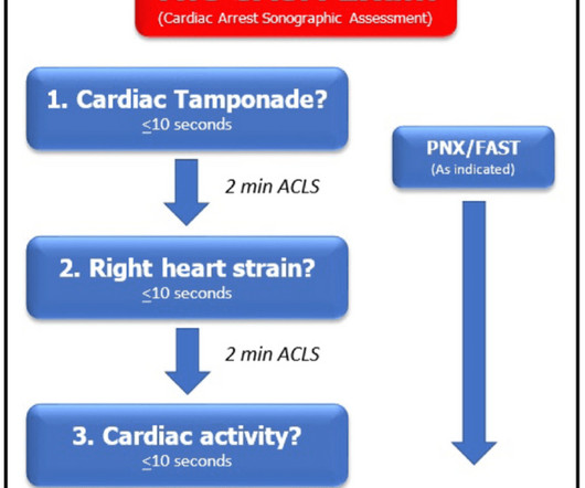
Ultrasound during cardiac arrest has quickly become standard. Initially, data suggested that the use of ultrasound during arrest increased pauses between compressions which worsens outcomes. The ideal view depends on the patient’s comorbid conditions such as COPD, obesity, cachexia, etc.

article thumbnail

SAEM Clinical Images Series: An Ultrasonographic Rabbit Hole

ALiEM

An 86-year-old man with a past medical history of coronary artery disease, hypertension, hyperlipidemia, chronic kidney disease, COPD, choledocholithiasis requiring ERCP and sphincterotomy 2 years ago presented with five days of feeling unwell. Gangrenous cholecystitis: diagnosis by ultrasound. Ultrasound, 30: 270-274.

Insiders

Sign Up for our Newsletter

This site is protected by reCAPTCHA and the Google Privacy Policy and Terms of Service apply.

article thumbnail

Consider The Probe: Spine Sign - It's Got Your Back!

Cook County EM Blog

The Case: A 68-year-old male with a history of CHF, COPD, CAD s/p stenting, HTN, and DM presents to the emergency department with worsening dyspnea and bilateral lower extremity edema for one week with associated orthopnea, and increased home oxygen requirement. Point of Care Ultrasound is shown below: Clip 1. VS: T 36.6, VS: T 36.6,

article thumbnail

Probing 2023: PoCUS Articles to Change Your Practice

EM Ottawa

Point-of-care ultrasound (PoCUS) is becoming a cornerstone tool in our assessment of patient presentations and is helping us guide our clinical management. This is also represented in the amount of literature that has been published on the use of PoCUS in 2023.

article thumbnail

emDOCs Podcast – Episode 98: Post ROSC Mental Model

EMDocs

Check the pulse RSI= Resuscitation Sequence Intubation Hypoxia, Hypotension, and Acidosis are the reason patients code during/post intubation These patients are super high risk for all 4 Optimize first pass success – Induction agent + paralytic Unconscious patients will still have muscle tone Induction Ketamine or Etomidate at half doses (i.e.,

article thumbnail

#FOAMed Review 49th Edition

EM Curious

EM Updates DVT ULTRASOUND DELUX [PODCAST]: Sometimes two point compression ultrasound may not be enough. Ultrasound Podcast brings us whole leg DVT ultrasound education.from a hotel room. FUEL FOR THE PE FIRE PE IN COPD: [BLOG]: How often do you think about PE in a clear COPD exacerbation?

COPD 52
article thumbnail

A 40-something woman with acute pulmonary edema -- see the Speckle Tracking echocardiogram.

Dr. Smith's ECG Blog

A 49 year old woman with h/o COPD only presented with sudden dyspnea. On arrival, lung ultrasound confirmed pulmonary edema (B lines). She had acute pulmonary edema on exam. There is STE and hyperacute T-waves in V2 and V3, with significant STE in I and aVL, and inferior reciprocal STD.

EKG/ECG 52