Remove COPD Remove Shock Remove Wellness
article thumbnail

ABG Versus VBG in the Emergency Department

EMDocs

Louis); Marina Boushra, MD (EM-CCM, Cleveland Clinic Foundation); Brit Long, MD (@long_brit) Case Emergency Medical Services brings in a 62-year-old male with COPD in acute on chronic hypoxemic respiratory failure (usually on 3 L nasal cannula, now on non-rebreather at 15 L/min). proning patients, ECMO) (12).

article thumbnail

ECG Blog #366 — Diltiazem didn't work.

Ken Grauer, MD

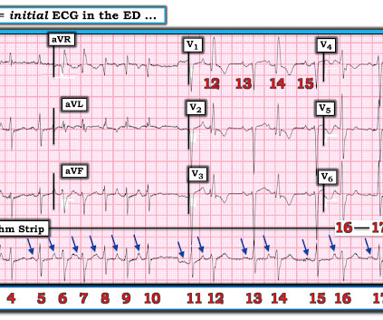
These 2 settings are: i ) In patients with severe , often longstanding pulmonary disease ; and / or , ii ) In acutely ill patients with multi-system disease ( ie, sepsis, shock, electrolyte and/or acid-base disorders ). Applying the Above to Today's Case: In addition to being Covid-positive — the patient in today's case had longstanding COPD.

EKG/ECG 195
Insiders

Sign Up for our Newsletter

This site is protected by reCAPTCHA and the Google Privacy Policy and Terms of Service apply.

Trending Sources

article thumbnail

Chest discomfort and a dilated right ventricle. What's going on?

Dr. Smith's ECG Blog

There is some upwards concave ST segment elevation in the inferior leads with what seems to be well formed J-waves. maybe not seen well on these echo-loops) The CW doppler at the tricuspid valve showed a maximum TR velocity of 2,55m/s with a TRP gradient of 26mmHg. Not much difference from 1st ECG. A bedside echo was performed.

EKG/ECG 99
article thumbnail

Diagnostics and Therapeutics: Managing Pneumothorax

Taming the SRU

This topic is additionally complicated by the development of multiple diagnostic tools now available for diagnosis as well as variable sizing algorithms used around the world. J Emerg Trauma Shock. Institutional resources and specialty services may further dictate the management of PTX. Ann Emerg Med. 1983;12(6):411-412.

article thumbnail

POCUS findings of hemodynamically unstable PE with cardiac arrest

EMDocs

RV chamber size alone is not enough information to rule-in a PE as RV cavity enlargement can be visualized in other conditions such as pulmonary hypertension, RV infarct, COPD and cardiac arrest from multiple causes. If RV pressure is extreme, it can exceed LV pressure causing paradoxical septal wall motion toward the LV as well.

article thumbnail

A crashing patient with an abnormal ECG that you must recognize

Dr. Smith's ECG Blog

This includes, but is not limited to, PE, asthma/COPD exacerbation, hypoxic vasoconstriction from pneumonia, acute pulmonary hypertension exacerbation. When there is tachycardia, the patient is in cardiogenic shock with very poor LV function on bedside echo. She was discharged and did well. Here is an example of Wellens'.

EKG/ECG 52
article thumbnail

A woman in her 20s with syncope

Dr. Smith's ECG Blog

Given her tachycardia and episodes of syncope, the patient was judged to be in compensated obstructive shock with very high risk of imminent decompensation. There appears to be some ST elevation in leads II and aVF, as well as to a lesser extent in the lateral chest leads. Thus, there is diffuse low voltage.

EKG/ECG 52