Remove COPD Remove Outcomes Remove Shock
article thumbnail

Serial PoCUS for ED Patients with Acute Dyspnea: Is More Actually Better?

RebelEM

It can be used to distinguish between various conditions, including chronic obstructive pulmonary disease (COPD) exacerbation, acute heart failure (AHF), pleural effusion, pulmonary edema, pericardial effusion, pneumothorax, and pneumonia [2,3]. Patients : Compared standard of care to serial US plus stand care in patients with dyspnea.

article thumbnail

ECG Blog #366 — Diltiazem didn't work.

Ken Grauer, MD

These 2 settings are: i ) In patients with severe , often longstanding pulmonary disease ; and / or , ii ) In acutely ill patients with multi-system disease ( ie, sepsis, shock, electrolyte and/or acid-base disorders ). Applying the Above to Today's Case: In addition to being Covid-positive — the patient in today's case had longstanding COPD.

EKG/ECG 195
Insiders

Sign Up for our Newsletter

This site is protected by reCAPTCHA and the Google Privacy Policy and Terms of Service apply.

Trending Sources

article thumbnail

Episode 21- Updates and Controversies in the Early Management of Sepsis and Septic Shock

EB Medicine

This month, we’ll be talking Updates and Controversies in the Early Management of Sepsis and Septic Shock. And sepsis-3 redefined septic shock as “hypotension not responsive to fluid resuscitation” with the added requirement of vasopressors to maintain a MAP greater than or equal to 65 and with a lactate > 2. That’s where we’re at.

Sepsis 40
article thumbnail

Diagnostics and Therapeutics: Managing Pneumothorax

Taming the SRU

It can be further divided into two types: primary--those that occur in generally healthy individuals without underlying lung disease, and secondary--those that occur in individuals with underlying lung disease such as COPD [1]. J Emerg Trauma Shock. Persistent air-leak in spontaneous pneumothorax--clinical course and outcome.

article thumbnail

Chest discomfort and a dilated right ventricle. What's going on?

Dr. Smith's ECG Blog

COPD, Idiopathic PAH, acute or chronic PE, pulmonary valve stenosis, etc) 3) Conditions affecting RV myocardial contractility, such as ARVD or RV infarction The ECGs does not really show any signs of chronic RV dilation or hypertrophy. Any cause of pulmonary hypertension. Our THANKS to Dr. Magnus Nossen for sharing this case with us.

EKG/ECG 94
article thumbnail

ABG Versus VBG in the Emergency Department

EMDocs

Louis); Marina Boushra, MD (EM-CCM, Cleveland Clinic Foundation); Brit Long, MD (@long_brit) Case Emergency Medical Services brings in a 62-year-old male with COPD in acute on chronic hypoxemic respiratory failure (usually on 3 L nasal cannula, now on non-rebreather at 15 L/min). is more likely to portend death (13, 14).

article thumbnail

What I read last week 9/22/24

Sensible Medicine

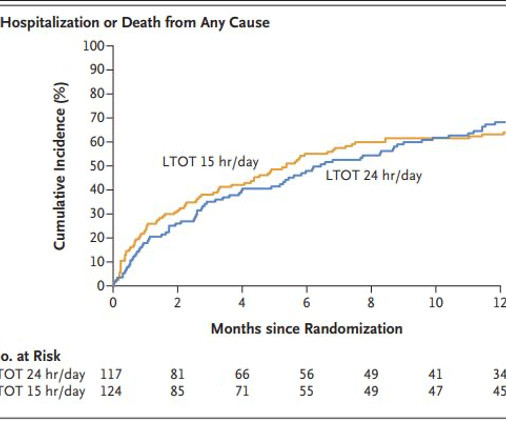
In studies, and in practice, these people tend to be patients with COPD who have resting O 2 sats of < 88%. The primary outcome was a composite of hospitalization or death from any cause within one year. About 80% had either COPD or pulmonary fibrosis and the mean PaO 2 and O 2 sats were about 50mm Hg and 80%, respectively.

COPD 86