Remove COPD Remove EKG/ECG Remove Wellness
article thumbnail

ECG Blog #366 — Diltiazem didn't work.

Ken Grauer, MD

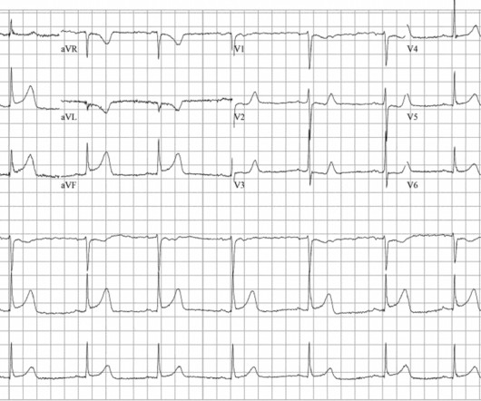
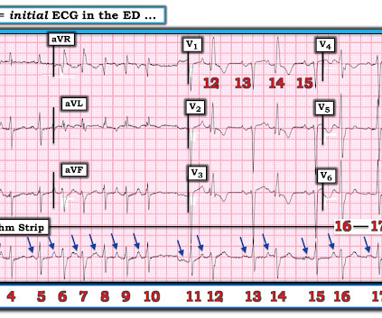
The ECG and long lead II rhythm strip in Figure-1 — was obtained from a COVID positive patient with persistent tachycardia not responding to Diltiazem. Figure-1: The initial ECG — obtained from a patient with persistent tachycardia. ( To improve visualization — I've digitized the original ECG using PMcardio ).

EKG/ECG 195
article thumbnail

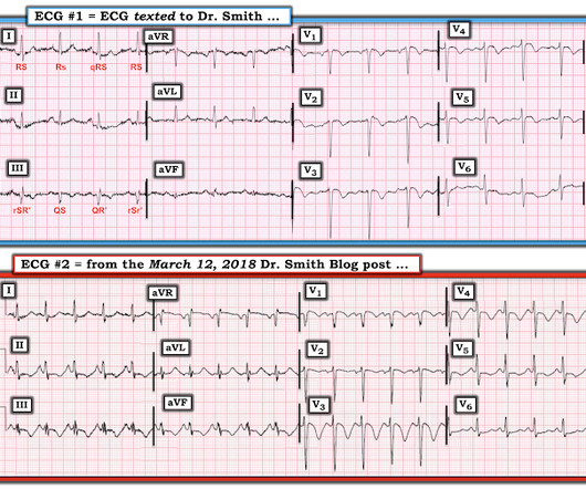
Proportionality is a major element in the ECG Diagnosis of OMI.

Dr. Smith's ECG Blog

This is the result for this ECG, from MDcalc.com : The most accurate cutpoint is 18.2. And she learned it well: She is not highly confident, but she does diagnose OMI. Here is a similar case from Pendell: This ECG was handed over at triage. A value above 18.2 (LAD A value above 19 is about 97% specific for LAD OMI.

EKG/ECG 110
Insiders

Sign Up for our Newsletter

This site is protected by reCAPTCHA and the Google Privacy Policy and Terms of Service apply.

Trending Sources

article thumbnail

An elderly male with acute altered mental status and huge ST Elevation

Dr. Smith's ECG Blog

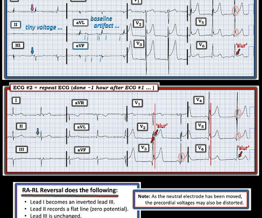
A man in his 90s with a history of HTN, CKD, COPD, and OSA presented to the emergency department after being found unresponsive at home. EKG on arrival to the ED is shown below: What do you think? ng/mL (consistent with prior baseline), and a repeat EKG was obtained 1 hour after the initial EKG. or basilar ischemia.

EKG/ECG 95
article thumbnail

Case Report: Coronary Vasospasm-Induced Cardiac Arrest

ACEP Now

A 45-year-old male with a history of chronic obstructive pulmonary disease (COPD), asthma, amphetamine and tetrahydrocannabinol (THC) use, and coronary vasospasm presented to triage with chest pain. During initial assessment, an ECG was obtained and revealed ST-segment elevation (STE) in the inferior leads with ST depression anteriorly.

article thumbnail

Chest discomfort and a dilated right ventricle. What's going on?

Dr. Smith's ECG Blog

The following ECG was recorded. The presenting ECG shows SR with narrow QRS complexes. There is some upwards concave ST segment elevation in the inferior leads with what seems to be well formed J-waves. First troponin I returned 3174 ng/l, at which point a repeat ECG was ordered. Not much difference from 1st ECG.

EKG/ECG 94
article thumbnail

Massive Hemoptysis

EM SIM Cases

He previously served as Deputy Medical Director at Andersen Simulation Center, the largest simulation center in the US Army as well as the Simulation Director for Madigan Army Medical Center’s Department of Emergency Medicine Residency. His initial presentation will respond to traditional therapies for COPD exacerbation.

COPD 52
article thumbnail

What is lurking underneath this new right bundle branch block?

Dr. Smith's ECG Blog

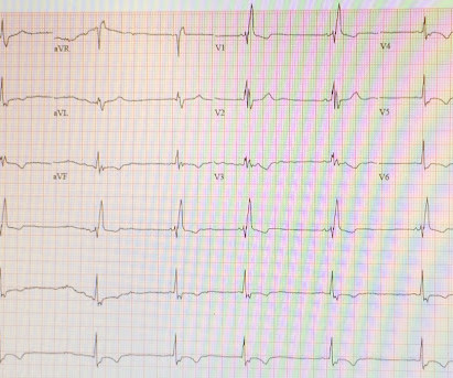
Written by Pendell Meyers, edits by Smith: Case A 72 year old female with hypertension and COPD presented with sudden shortness of breath and chest pain. Here is her triage ECG (the baseline is not available but reportedly "normal"): What is your interpretation? There is sinus rhythm with PACs and PVCs. Learning Points: 1.

COPD 52