Remove COPD Remove EKG/ECG Remove Shock
article thumbnail

ECG Blog #366 — Diltiazem didn't work.

Ken Grauer, MD

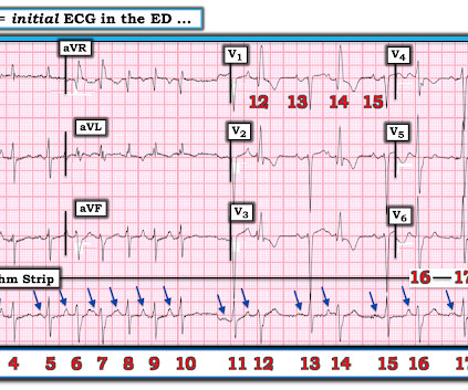
The ECG and long lead II rhythm strip in Figure-1 — was obtained from a COVID positive patient with persistent tachycardia not responding to Diltiazem. Figure-1: The initial ECG — obtained from a patient with persistent tachycardia. ( To improve visualization — I've digitized the original ECG using PMcardio ).

EKG/ECG 195
article thumbnail

Chest discomfort and a dilated right ventricle. What's going on?

Dr. Smith's ECG Blog

The following ECG was recorded. The presenting ECG shows SR with narrow QRS complexes. First troponin I returned 3174 ng/l, at which point a repeat ECG was ordered. Not much difference from 1st ECG. At this point an old ECG on file was found for comparison. Upon admission she had ongoing slight chest discomfort.

EKG/ECG 103
Insiders

Sign Up for our Newsletter

This site is protected by reCAPTCHA and the Google Privacy Policy and Terms of Service apply.

article thumbnail

Valvular Emergencies

EB Medicine

Aortic Valve Disease Types and causes Mitral Valve Disease Types and causes Tricuspid and Pulmonic Disease Differential Diagnosis acute coronary syndromes (ACS) pulmonary embolism tamponade chronic obstructive pulmonary disease (COPD) pneumonia pneumothorax Prehospital Care Evaluation of chest pain History Shock ED Evaluation History Aortic Stenosis (..)

COPD 52
article thumbnail

Diffuse Subendocardial Ischemia on the ECG. Left main? 3-vessel disease? No!

Dr. Smith's ECG Blog

It was edited by Smith CASE : A 52-year-old male with a past medical history of hypertension and COPD summoned EMS with complaints of chest pain, weakness and nausea. An immediate 12-lead EKG was obtained: There is ST elevation in leads aVR and V1, with marked ST depression in I, II, III, aVF, V3-V6. What should be done?

EKG/ECG 40
article thumbnail

A crashing patient with an abnormal ECG that you must recognize

Dr. Smith's ECG Blog

Written by Pendell Meyers, with edits from Steve Smith Let's consider this nearly pathognomonic ECG without the clinical context (because sometimes the clinical context will not be as easy as in this case). This ECG is diagnostic of hemodynamically significant acute right heart strain. What is the answer? Why is it not Wellens???

EKG/ECG 40
article thumbnail

Are these Wellens' waves?

Dr. Smith's ECG Blog

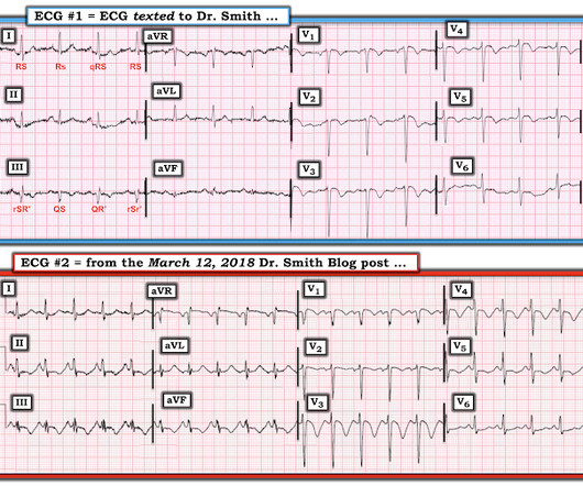
An ECG was texted to me (Smith) without any clinical information: What did I say? This clinical information followed: "The patient had a COPD exacerbation with a prehospital SpO2 of 60%. This clinical information followed: "The patient had a COPD exacerbation with a prehospital SpO2 of 60%. This is NOT Wellens.

EKG/ECG 52
article thumbnail

Atrial fibrillation? Multifocal Atrial Tachycardia? Don't look at computer read until AFTER you interpret!

Dr. Smith's ECG Blog

This 60-something with h/o COPD and HFrEF (EF 25%) presented with SOB and chest pain. Here is the ECG: What do you think? Shah and Rubin studied the computer rhythm interpretation of 2160 12-lead ECGs, compared to 2 cardiologists [ 18 ]. noted computer over-interpretation (false positives) of AF in 9 to 19% of ECGs [ 19 ].

EKG/ECG 52