Remove COPD Remove EKG/ECG Remove Sepsis
article thumbnail

ECG Blog #366 — Diltiazem didn't work.

Ken Grauer, MD

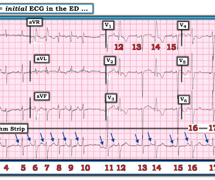
The ECG and long lead II rhythm strip in Figure-1 — was obtained from a COVID positive patient with persistent tachycardia not responding to Diltiazem. Figure-1: The initial ECG — obtained from a patient with persistent tachycardia. ( To improve visualization — I've digitized the original ECG using PMcardio ).

EKG/ECG 195
article thumbnail

emDOCs Podcast – Episode 98: Post ROSC Mental Model

EMDocs

Check the pulse RSI= Resuscitation Sequence Intubation Hypoxia, Hypotension, and Acidosis are the reason patients code during/post intubation These patients are super high risk for all 4 Optimize first pass success – Induction agent + paralytic Unconscious patients will still have muscle tone Induction Ketamine or Etomidate at half doses (i.e.,

Insiders

Sign Up for our Newsletter

This site is protected by reCAPTCHA and the Google Privacy Policy and Terms of Service apply.

article thumbnail

Atrial fibrillation? Multifocal Atrial Tachycardia? Don't look at computer read until AFTER you interpret!

Dr. Smith's ECG Blog

This 60-something with h/o COPD and HFrEF (EF 25%) presented with SOB and chest pain. Here is the ECG: What do you think? Shah and Rubin studied the computer rhythm interpretation of 2160 12-lead ECGs, compared to 2 cardiologists [ 18 ]. noted computer over-interpretation (false positives) of AF in 9 to 19% of ECGs [ 19 ].

EKG/ECG 52
article thumbnail

Diffuse Subendocardial Ischemia on the ECG. Left main? 3-vessel disease? No!

Dr. Smith's ECG Blog

It was edited by Smith CASE : A 52-year-old male with a past medical history of hypertension and COPD summoned EMS with complaints of chest pain, weakness and nausea. An immediate 12-lead EKG was obtained: There is ST elevation in leads aVR and V1, with marked ST depression in I, II, III, aVF, V3-V6. What should be done?

EKG/ECG 40