article thumbnail

ECG Blog #366 — Diltiazem didn't work.

Ken Grauer, MD

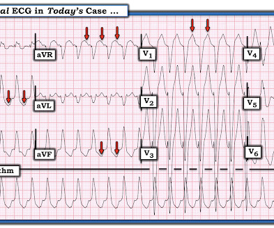
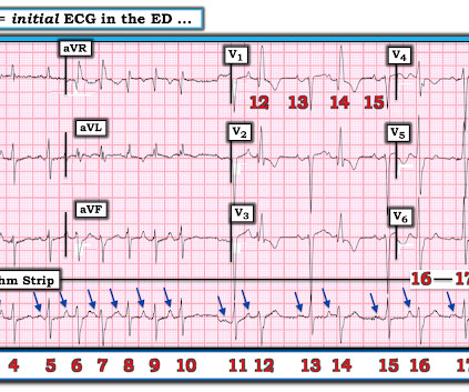
The ECG and long lead II rhythm strip in Figure-1 — was obtained from a COVID positive patient with persistent tachycardia not responding to Diltiazem. Figure-1: The initial ECG — obtained from a patient with persistent tachycardia. ( To improve visualization — I've digitized the original ECG using PMcardio ).

EKG/ECG 195
article thumbnail

VT? Or Supraventricular tachycardic rhythm with aberrancy?

Dr. Smith's ECG Blog

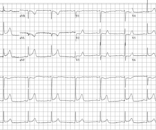
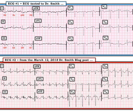
Here is a normal LBBB: In LBBB, monophasic wide R-waves should be limited to the lateral leads in left bundle branch block, as in this ECG. In that ECG above, there are monophasic R-waves starting in lead V2 all the way out to V6. But why does this EKG look nothing like left bundle branch block? Is this sinus tachycardia or VT?

EKG/ECG 140
Insiders

Sign Up for our Newsletter

This site is protected by reCAPTCHA and the Google Privacy Policy and Terms of Service apply.

article thumbnail

An elderly male with acute altered mental status and huge ST Elevation

Dr. Smith's ECG Blog

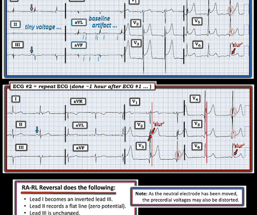
A man in his 90s with a history of HTN, CKD, COPD, and OSA presented to the emergency department after being found unresponsive at home. EKG on arrival to the ED is shown below: What do you think? ng/mL (consistent with prior baseline), and a repeat EKG was obtained 1 hour after the initial EKG. or basilar ischemia.

EKG/ECG 105
article thumbnail

Proportionality is a major element in the ECG Diagnosis of OMI.

Dr. Smith's ECG Blog

This is the result for this ECG, from MDcalc.com : The most accurate cutpoint is 18.2. Here is a similar case from Pendell: This ECG was handed over at triage. That said — the ST-T wave in lead V2 looks to be small only if viewed in the context of its absolute height, as one of 12 leads in the initial ECG shown above in today's post.

EKG/ECG 120
article thumbnail

emDOCs Podcast – Episode 98: Post ROSC Mental Model

EMDocs

Check the pulse RSI= Resuscitation Sequence Intubation Hypoxia, Hypotension, and Acidosis are the reason patients code during/post intubation These patients are super high risk for all 4 Optimize first pass success – Induction agent + paralytic Unconscious patients will still have muscle tone Induction Ketamine or Etomidate at half doses (i.e.,

article thumbnail

Is all this "ST Depression" due to ischemia?

Dr. Smith's ECG Blog

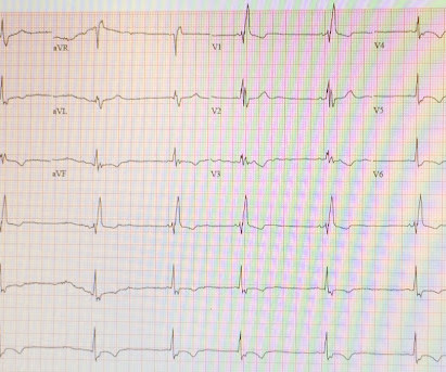
Written by Magnus Nossen, with some edits by Smith This ECG was transmitted electronically by EMS for evaluation. How would you interpret the ST changes seen in this ECG? Will you accept this patient for emergent coronary angiogram based on the ECG changes? Does the ECG represent STEMI-negative OMI findings?

EKG/ECG 77
article thumbnail

Chest discomfort and a dilated right ventricle. What's going on?

Dr. Smith's ECG Blog

The following ECG was recorded. The presenting ECG shows SR with narrow QRS complexes. First troponin I returned 3174 ng/l, at which point a repeat ECG was ordered. Not much difference from 1st ECG. At this point an old ECG on file was found for comparison. Upon admission she had ongoing slight chest discomfort.

EKG/ECG 103