Remove COPD Remove Documentation/Coding Remove Shock
article thumbnail

ECG Blog #366 — Diltiazem didn't work.

Ken Grauer, MD

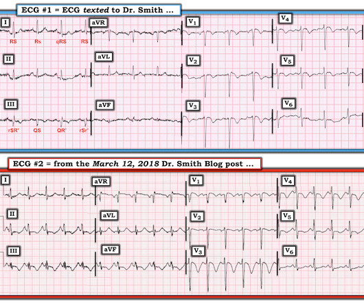
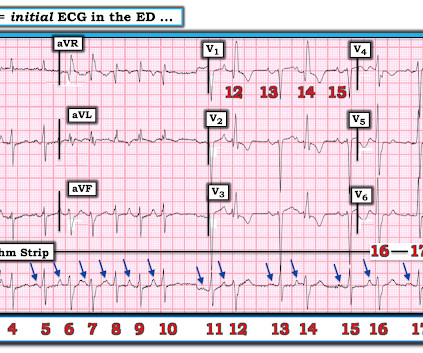
Figure-2: I've color-coded P waves from Figure-1 according to P wave morphology ( See text ). NOTE: For clarity — I've color-coded P waves in the long lead II rhythm strip according to morphology. Applying the Above to Today's Case: In addition to being Covid-positive — the patient in today's case had longstanding COPD.

EKG/ECG 195
article thumbnail

Episode 21- Updates and Controversies in the Early Management of Sepsis and Septic Shock

EB Medicine

This month, we’ll be talking Updates and Controversies in the Early Management of Sepsis and Septic Shock. And sepsis-3 redefined septic shock as “hypotension not responsive to fluid resuscitation” with the added requirement of vasopressors to maintain a MAP greater than or equal to 65 and with a lactate > 2. That’s where we’re at.

Sepsis 40
Insiders

Sign Up for our Newsletter

This site is protected by reCAPTCHA and the Google Privacy Policy and Terms of Service apply.

article thumbnail

Diffuse Subendocardial Ischemia on the ECG. Left main? 3-vessel disease? No!

Dr. Smith's ECG Blog

It was edited by Smith CASE : A 52-year-old male with a past medical history of hypertension and COPD summoned EMS with complaints of chest pain, weakness and nausea. Clinical Course The paramedic activated a “Code STEMI” alert and transported the patient nearly 50 miles to the closest tertiary medical center. What do you see?

EKG/ECG 40
article thumbnail

Are these Wellens' waves?

Dr. Smith's ECG Blog

This clinical information followed: "The patient had a COPD exacerbation with a prehospital SpO2 of 60%. Tachycardia is unusual in ACS unless there is cardiogenic shock or a second simultaneous pathology. Many patients with acute COPD exacerbations present with a similar heart rate. This is NOT Wellens. Is the patient hypoxic?

EKG/ECG 52
article thumbnail

Atrial fibrillation? Multifocal Atrial Tachycardia? Don't look at computer read until AFTER you interpret!

Dr. Smith's ECG Blog

This 60-something with h/o COPD and HFrEF (EF 25%) presented with SOB and chest pain. See below how this has been documented. Here is the ECG: What do you think? Computer interpretation is below. Providers FORGET to “ U se t he O dds”. Q UESTION : Is there a d ifference between sinus rhythm with multiple PACs vs MAT?

EKG/ECG 52
article thumbnail

A woman in her 20s with syncope

Dr. Smith's ECG Blog

Given her tachycardia and episodes of syncope, the patient was judged to be in compensated obstructive shock with very high risk of imminent decompensation. This patient was reported to have distant heart sounds but was not hypotensive and did not have JVD according to documentation.

EKG/ECG 40
article thumbnail

ABG Versus VBG in the Emergency Department

EMDocs

Louis); Marina Boushra, MD (EM-CCM, Cleveland Clinic Foundation); Brit Long, MD (@long_brit) Case Emergency Medical Services brings in a 62-year-old male with COPD in acute on chronic hypoxemic respiratory failure (usually on 3 L nasal cannula, now on non-rebreather at 15 L/min). is more likely to portend death (13, 14).