article thumbnail

ECG Blog #389 — A Quote from Sherlock Holmes

Ken Grauer, MD

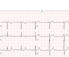
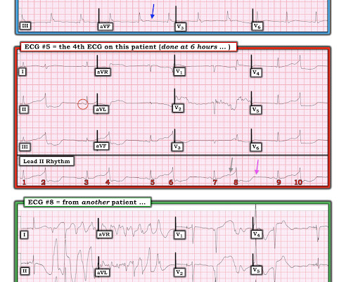
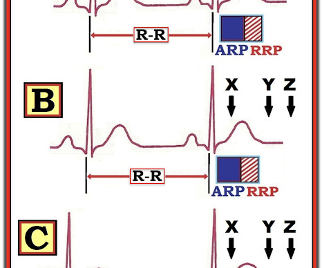
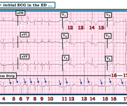
MY Approach to the Rhythm in Figure-1: As per ECG Blog #185 — I favor the P s, Q s, 3 R Approach for interpretation of the cardiac rhythm — beginning with whichever of these 5 KEY Parameters is easiest to assess for the tracing in front of me: At least in the single lead II rhythm strip seen in Figure-1 — The Q RS complex appears to be narrow.

EKG/ECG 370
article thumbnail

Chest Pain in Children: ReBaked Morsel

Pediatric EM Morsels

EKG Reasonable screen for cardiac etiology [ Kane, 2010 ]: Chest Pain with Exertion? The role of point-of-care ultrasound in the diagnosis of pericardial effusion: a single academic center retrospective study. Is there a family history of sudden death, hypertrophic cardiomyopathy, long QT syndrome, or tachyarrhythmias? Ultrasound J.

EKG/ECG 271
Insiders

Sign Up for our Newsletter

This site is protected by reCAPTCHA and the Google Privacy Policy and Terms of Service apply.

article thumbnail

ECG Blog #395 — No Simple Explanation.

Ken Grauer, MD

Regular followers of my ECG Blog know I routinely emphasize a series of user - friendly points to facilitate the interpretation of any rhythm. L addergram I llustration : Academically — I was fascinated by today's rhythm. But at this point, since I could not yet explain all ECG features — I needed to draw a laddergram.

EKG/ECG 245
article thumbnail

ECG Blog #371 — Palpitations Since Childhood.

Ken Grauer, MD

The ECG in Figure-1 is from a man in his 30s — who overall has been healthy, except for a history of "intermittent palpitations" that he has had since childhood. He was hemodynamically stable with ECG #1. Figure-1: The initial ECG in today's case. To improve visualization — I've digitized the original ECG using PMcardio ).

EKG/ECG 195
article thumbnail

Occlusion myocardial infarction is a clinical diagnosis

Dr. Smith's ECG Blog

Willy is a cardiology fellow with a keen interest in the ECG in OMI. If an immediate EKG was obtained, it was not saved in the medical record. The first available EKG was recorded just after midnight, presumably around the time the result of the troponin came to clinical attention. The patient said, "I just don't feel good."

EKG/ECG 124
article thumbnail

Very fast regular tachycardia: 2 ECGs from the same patient. What is going on?

Dr. Smith's ECG Blog

An ECG was recorded immediately and is shown below. How do you interpret the ECG? ECG#1 There is a regular tachycardia with a ventricular rate of about 180 bpm. After cardioversion, if successful, you can take a few moments to assess the 12-lead in more detail and assess the post conversion ECG. ECG#2 What do you think?

EKG/ECG 134
article thumbnail

Sudden shock with a Nasty looking ECG. What is it?

Dr. Smith's ECG Blog

There were 2 prehospital ECGs: What do you think? When I was shown this ECG, I said it looks like such widespread ischemia that is might be a left main occlusion, or LM ischemia plus circumflex occlusion (high lateral and posterior OMI). Total Left Main Occlusion presents with different ECG findings which are multi-faceted.

Shock 128