Remove 2023 Remove EKG/ECG Remove Ultrasounds
article thumbnail

Concerning EKG with a Non-obstructive angiogram. What happened?

Dr. Smith's ECG Blog

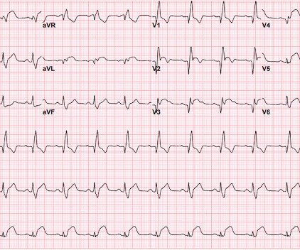
It appears EMS obtained two EKGs, but unfortunately these were not saved in the medical record. The EMS crew was only BLS certified, so EKG interpretation is not within their scope of practice. The patient arrived just after 10 AM, and the following EKG was obtained. There are no further EKGs or troponin measurements.

EKG/ECG 123
article thumbnail

Acute chest pain and an abnormal ECG. Do precordial leads show benign T-wave inversion or ischemia?

Dr. Smith's ECG Blog

ECG 1 What do you think? Benign T-wave Inversion -- Countless Examples (1) The Queen of Hearts Active OMI model (aOMI) is shown below: Willy : My initial impression looking at this ECG was that it was not ischemic. He concluded by saying that "History and concern should win regardless of EKG." It is imperfect.

EKG/ECG 75
Insiders

Sign Up for our Newsletter

This site is protected by reCAPTCHA and the Google Privacy Policy and Terms of Service apply.

article thumbnail

Another deadly triage ECG missed, and the waiting patient leaves before being seen. What is this nearly pathognomonic ECG?

Dr. Smith's ECG Blog

Pain improved to 1/10 after EMS administers 324 mg aspirin and the following EKG is obtained at triage. If this EKG were handed to you to screen from triage without any clinical information, what would you think? Do you appreciate any dynamic changes compared to the patient’s prior EKG? What do you think? In fact, Kosuge et al.

EKG/ECG 138
article thumbnail

Expert human ECG interpretation and/or the Queen of Hearts could have saved this patient's anterior wall

Dr. Smith's ECG Blog

He had active chest pain at the time of triage at 0137 at night, with this triage ECG: What do you think? I sent this ECG, without any text at all, to Dr. Smith, and he replied: "LAD OMI with low certainty. Back to the case: Unfortunately, the ECG was not understood by the provider. V3 is the one that is convincing."

EKG/ECG 127
article thumbnail

Dynamic OMI ECG. Negative trops and negative angiogram does not rule out coronary ischemia or ACS.

Dr. Smith's ECG Blog

Here is his ED ECG at triage: Obvious high lateral OMI that does not quite meet STEMI criteria. He had a previous ECG on file: Proving the findings are new The cath lab was activated. Bedside cardiac ultrasound with no obvious wall motion abnormalities. Bedside cardiac ultrasound with no obvious wall motion abnormalities.

EKG/ECG 119
article thumbnail

Four patients with chest pain and ‘normal’ ECG: can you trust the computer interpretation?

Dr. Smith's ECG Blog

All initial ECGs were labeled ‘normal’ or ‘otherwise normal’ by the computer interpretation, and below are the ECGs with the final cardiology interpretation. 1-3] But these studies were very short duration and used cardiology interpretation of ECGs or emergent angiography rather than patient outcomes.

EKG/ECG 119
article thumbnail

Acute artery occlusion -- which one?

Dr. Smith's ECG Blog

Her ECG is shown below: What do you think? The conventional machine algorithm interpreted this ECG as STEMI. Alternatively, with STE in V1 and III, and STD in I and aVL, this ECG could represent proximal RCA OMI with right ventricular involvement. What do you do clinically when the ECG looks like this?

EKG/ECG 115