Remove 2023 Remove EKG/ECG Remove Resuscitation
article thumbnail

ECG Blog #410 — How Tall are the T Waves?

Ken Grauer, MD

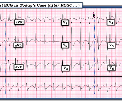
QUESTION: HOW would YOU interpret the ECG in Figure-1 — if no clinical information was provided? Figure-1: The initial ECG in today's case. ( To improve visualization — I've digitized the original ECG using PMcardio ). = The ECG in Figure-1 — was obtained following successful resuscitation.

EKG/ECG 434
article thumbnail

ECG Blog #415 — The Cath showed NO Occlusion!

Ken Grauer, MD

The ECG in Figure-1 was obtained following successful resuscitation. Stat Echo — obtained shortly after successful resuscitation revealed anterior wall akinesis. QUESTIONS: In view of the above history — How would YOU interpret the ECG in Figure-1 ? Is this ECG finding present in today’s initial ECG?

EKG/ECG 408
Insiders

Sign Up for our Newsletter

This site is protected by reCAPTCHA and the Google Privacy Policy and Terms of Service apply.

Trending Sources

article thumbnail

ECG Blog #388 — Why Does Lead V1 Look Funny?

Ken Grauer, MD

The ECG in Figure-1 was obtained from an 18-year old woman — who moments before been resuscitated from out-of-hospital cardiac arrest. How would YOU interpret her post-resuscitation ECG? Does this ECG in Figure-1 provide clue(s) to the etiology of this patient's cardiac arrest? About A RVC/ A RVD.

EKG/ECG 370
article thumbnail

ECG Blog #448 — A Young Man with Chest Pain.

Ken Grauer, MD

The ECG in Figure-1 was obtained from a previously healthy man in his early 20s — who initially presented with GI symptoms, that then evolved into CP ( C hest P ain ). QUESTIONS: Given the above history — How would YOU interpret the initial ECG that is shown in Figure-1 ? Figure-1: The initial ECG in today's case. (

EKG/ECG 300
article thumbnail

ECG Blog #389 — A Quote from Sherlock Holmes

Ken Grauer, MD

MY Approach to the Rhythm in Figure-1: As per ECG Blog #185 — I favor the P s, Q s, 3 R Approach for interpretation of the cardiac rhythm — beginning with whichever of these 5 KEY Parameters is easiest to assess for the tracing in front of me: At least in the single lead II rhythm strip seen in Figure-1 — The Q RS complex appears to be narrow.

EKG/ECG 370
article thumbnail

ECG Blog #401 — What Kind of Block?

Ken Grauer, MD

The ECG in Figure-1 was obtained from an elderly woman — who presented to the ED ( E mergency D epartment ) for dyspnea on exertion over recent weeks. Figure-1: The initial ECG in today's case. ( To improve visualization — I've digitized the original ECG using PMcardio ). What are YOUR "Quick Thoughts" about this case?

EKG/ECG 254
article thumbnail

ECG Blog #364 — VT in Need of Cardioversion?

Ken Grauer, MD

T he ECG in Figure-1 was obtained from a 60ish year old man on arrival to the ED ( E mergency D epartment ). Figure-1: The initial ECG in today's case — obtained from a 60ish year old man who collapsed in the ED shortly after this tracing was recorded. ( To improve visualization — I've digitized the original ECG using PMcardio ).

EKG/ECG 195