This site uses cookies to improve your experience. To help us insure we adhere to various privacy regulations, please select your country/region of residence. If you do not select a country, we will assume you are from the United States. Select your Cookie Settings or view our Privacy Policy and Terms of Use.
Cookie Settings
Cookies and similar technologies are used on this website for proper function of the website, for tracking performance analytics and for marketing purposes. We and some of our third-party providers may use cookie data for various purposes. Please review the cookie settings below and choose your preference.
Used for the proper function of the website
Used for monitoring website traffic and interactions
Cookie Settings
Cookies and similar technologies are used on this website for proper function of the website, for tracking performance analytics and for marketing purposes. We and some of our third-party providers may use cookie data for various purposes. Please review the cookie settings below and choose your preference.
Strictly Necessary: Used for the proper function of the website
Performance/Analytics: Used for monitoring website traffic and interactions
Ultrasound can assist: confirm ascites, evaluate for best site, abdominal wall thickness, blood vessels along needle track. Management: Patients can rapidly progress to septic shock and multiorgan failure. Safety of ultrasound-guided thoracentesis in patients with abnormal preprocedural coagulation parameters. 2013;144:456–463.
Ischemic Hepatitis and Septic Shock Secondary to Murine Typhus Infection in Pregnancy. The RUSH exam: Rapid Ultrasound in SHock in the evaluation of the critically lll. January 2022. Answer : Murine Typhus Epidemiology: Murine typhus is endemic in several parts of the U.S.: Clinical Infectious Diseases , vol. 6, 2008, pp.
A 50-something man presented in shock with severe chest pain. The patient was in clinical shock with a lactate of 8. This confirms inferior, posterior, lateral, and RV MI RV MI often leads to shock and (systolic) hypotension. Case continued A bedside ultrasound showed diminished LV EF and of course bradycardia.
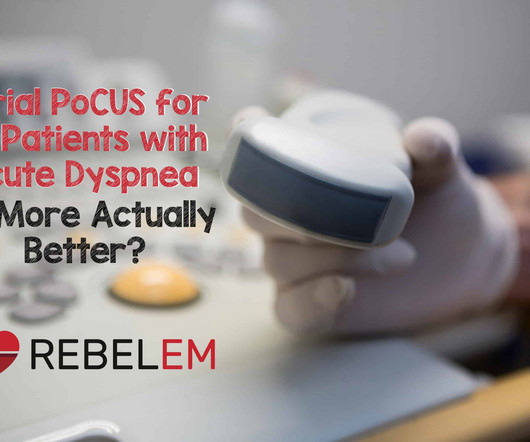
Background: Point-of-care ultrasound (PoCUS) is a valuable clinical tool in the assessment of acute dyspnea. Impact of serial cardiopulmonary point-of-care ultrasound exams in patients with acute dyspnoea: a randomized, controlled trial. PoCUS evaluations included lung ultrasound (LUS) and focused cardiac ultrasound (FoCUS).
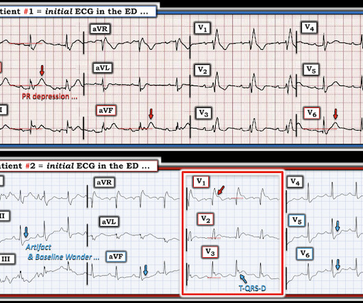
Taking a step back , remember that sinus tachycardia is less commonly seen in OMI (except in cases of impending cardiogenic shock). Answer : Bedside ultrasound! Smith : RV infarct may also have this appearance on ultrasound. So hypoxia without B lines on lung ultrasound strongly weights toward PE. Both were wrong.
Shocked x 2 without effect. Pads were placed with ultrasound guidance, so they were in the correct position. Warning: if this is VT, a calcium channel blocker can result in shock and death. We are told that " the Patient was Shocked X2 Without Effect." However, this is not SVT. What to do now? K returned 3.6
’ This could be done with the Venous Excess Ultrasound Score [VExUS] [5, 6]. Beaubien-Souligny W, Rola P, Haycock K, Bouchard J, Lamarche Y, Spiegel R et al : Quantifying systemic congestion with Point-Of-Care ultrasound: development of the venous excess ultrasound grading system. The ultrasound journal 2020, 12(1):1-12.
Point-of-Care-Ultrasound (POCUS) is a bedside modality that can assist Emergency Physicians (EPs) in differentiating PE from other causes of cardiac arrest. Multiorgan POCUS The diagnostic power of POCUS often resides in combining multiple ultrasound exams. 1-3 As many as 25% of acute PE cases present as sudden cardiac death.
The current standard of practice has moved away from landmark-based central line placement given the efficacy and safety of ultrasound-based techniques. 2022 PMID: 35206981 2. Healthcare 2022 PMID: 35206981 Post Peer Reviewed By: Frank Lodeserto, MD & Salim R. minutes compared to a mean placement time of 10.7 Healthcare.
R4 Case Follow-up: SCAD WITH dr. Martella Spontaneous Coronary Artery Dissection (SCAD) is a diagnosis confirmed via imaging: Coronary Angiography, Optical Coherence Tomography, Intravascular Ultrasound Therefore, treatment in the ED is the same as atherosclerotic ACS: ASA, heparin gtt and possible statin.
POCUS: RUSH exam (Rapid Ultrasound for Shock and Hypotension) While derived to help identify unexplained hypotension, if we accept that sinus tachycardia may be an early indicator of shock, we can utilize this general approach to help reevaluate as clinically indicated. Axilla, Sacral, GU Overall Volume Status? In: StatPearls.
Clinical impact: The patient’s DVT ultrasounds were negative. Drug Checking Quarterly Report (Q3 2022). 2022; 06/26/2023. 2022 May;14(5). Evaluation for nephrotic syndrome due to viral hepatitis, endocarditis, venous thromboembolism or other cause of chronic lymphedema is reasonable. 2011 Dec;6:1-4.
VF/asystole), a pulse cannot be identified via Doppler ultrasound for a full minute, or if lack of organized cardiac activity is confirmed on bedside echocardiogram. This is a good time to utilize an arterial line or use the ultrasound to find a central pulse. Published 2022 Jan 3. doi.org/10.1016/j.resuscitation.2018.11.004
International Consensus Criteria for Pediatric Sepsis and Septic Shock. The aim of this paper was to update and evaluate the criteria for sepsis and septic shock in children. Check out DFTB’s module on SIRS, Sepsis and Shock Module – Don’t Forget the Bubbles (dontforgetthebubbles.com) Why does it matter?
Follow this algorithm in patients with unstable bradycardia with acute heart failure, change in mental status, or concern for shock, physicians should start with atropine, 1 mg and may be continued every 3 to 5 minutes if effective. The American Journal of Emergency Medicine , vol. 2090–2093, [link] Moayedi, Siamak, et al. Circulation , vol.
Diagnosis because P ra is non-invasively transduced with ultrasound, for instance via the size and collapsibility of the inferior vena cava [1] and/or the Venous Excess Ultrasound Score [VExUS] [2-4]. If changes in P ra cause changes in CO, shouldn’t we then know the etiology of shock with certainty?
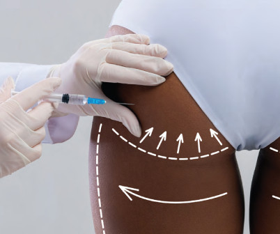
Unfortunately, at least 13 deaths were subsequently reported in Miami-Dade County from 2019–2022, the three years immediately following the implementation of the subcutaneous injection regulation. 6 Theoretically, ultrasound guidance should decrease this risk, which formed the basis for the 2022 Florida Medical Board regulation.
doi:10.1056/NEJMoa2212663 BACKGROUND Sepsis, including severe sepsis and septic shock, is a frequently encountered condition in the emergency department and carries a high mortality rate. 2022), ideally with antibiotics administered within one hour of sepsis recognition by the treating provider (Evans, Rhodes et al. N Engl J Med.
The patient in today’s case presented in cardiogenic shock from proximal LAD occlusion, in conjunction with a subtotally stenosed LMCA. Another approach is sympathetic chain (stellate ganglion) blockade if you have the skills to do it: it requires some expertise and ultrasound guidance. RCA — 100% proximal occlussion.
A recent observational study was performed to pragmatically assess clinically meaningful differences in BP in a diverse critically ill cohort with shock. In general, radial artery readings in patients with shock likely underestimate central pressure which can lead to increasing vasopressor dosing. 2022 Jul;45(7):1183-1192.
As of 2022, 1 in 4 emergency departments in the United States were staffed by a private equity-owned physician group. He was shocked when the hospital CEO took him up on it, but a year later, volumes increased 30 percent with his emphasis on quality rather than cost-cutting. 2022:3(9):e222886. Published December 14, 2022.
The ATHOS-3 trial in 2017 explored the efficacy of angiotensin II as a vasopressor for severe vasodilatory shock. Severe shock is defined as persistent hypotension requiring vasopressors to maintain a mean arterial pressure of 65mmHg and serum lactate <2 despite adequate volume resuscitation. were more likely to respond.
Example of pneumoperitoneum following perforated NEC Other Investigations Blood tests and abdominal ultrasound. Abdominal ultrasound may show bowel wall thickening, altered vascular state and free fluid. Updated 2022 Aug 8]. Treasure Island (FL): StatPearls Publishing; 2022 Jan-. Necrotizing Enterocolitis. Lakhoo, K. &
Possibly more useful in trauma patients during e-FAST to determine presence or absence of PTX and less helpful in spontaneous PTX where management is partially based on size, which is difficult to quantify on ultrasound and may be falsely positive in the setting of emphysematous blebs. J Emerg Trauma Shock. Ann Emerg Med. Can Respir J.
A 30-something woman with chest pain and h/o pulmonary hypertension due to chronic pulmonary emboli A 30-something with 8 hours of chest pain and an elevated troponin Syncope, Shock, AV block, Large RV, "Anterior" ST Elevation. Cardiac Ultrasound may be a surprisingly easy way to help make the diagnosis Answer: pulmonary embolism.
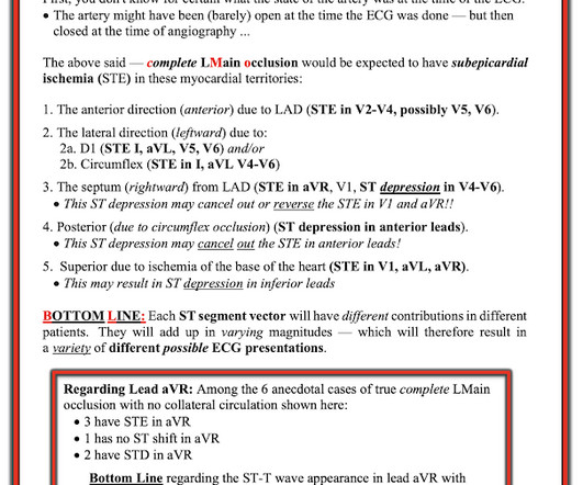
An elderly man with sudden cardiogenic shock, diffuse ST depressions, and STE in aVR Literature 1. Routine STEMI activation in STE-aVR for emergent revascularization is not warranted, although urgent, rather than emergent, catheterization appears to be important. == MY Comment, by K EN G RAUER, MD ( 11/4 /2022 ): == Our thanks to Drs.
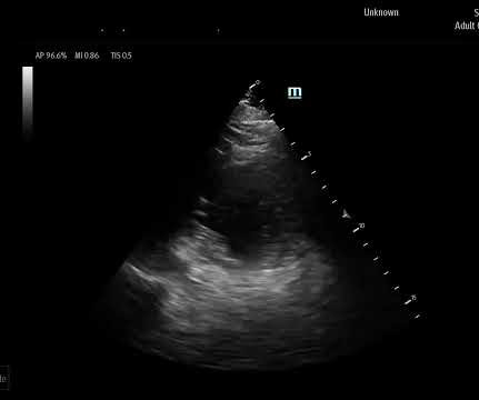
It was notable for a normal cardiac ultrasound with no pericardial fluid, normal LV and RV function (though the quality was not sufficient to evaluate for wall motion abnormalities) and normal IVC dynamics. Bedside ultrasound is another very important piece. Ultrasound can be very helpful to distinguish causes of hypotension.
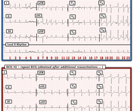
PMID: 34775811; PMCID: PMC9075358 A bedside ultrasound was performed, shown here: Parasternal short axis view demonstrating inferior LV wall motion akinesis Apical 2 chamber view again demonstrating inferior LV wall akinesis The cath lab was not activated based on the ECG and bedside echo. J Am Heart Assoc. 2021 Dec 7;10(23):e022866.
I would do bedside ultrasound to look at the RV, look for B lines as a cause of hypoxia (which would support OMI, and argue against PE), and if any doubt persists, a rapid CT pulmonary angiogram. As for the ECG, it could represent OMI, but RBBB is also a clue that it may be PE. There is sinus tachycardia at ~100/minute.
Tachycardia is unusual for OMI, unless the patient is in cardiogenic shock (or getting close). A bedside ultrasound should be done to assess volume and other etiologies of tachycardia, but if no cause of type 2 MI is found, the cath lab should be activated NOW. The October 21, 2022 post — for " artifactual VT".
Their argument required high levels of trust in measurements from ultrasound images. The first ORBITA trial shocked the cardiology world when it failed to find a placebo-resistant effect of stenting substantial coronary lesions. In 2022, I authored an opinion on AF screening in the New England Journal of Medicine.
A larger 2022 meta-analysis in the journal of Clinical Microbiology and Infection examined different test modalities for diagnosing pneumonia in the setting of clinical suspicion for acute community-acquired lower RTI in outpatient clinics, LTACs, and the emergency department. 2022 Jul 11;11(14):2163. 2022 Jan;28(1):13-22.
Her bedside cardiac ultrasound was normal We decided to cardiovert her since the time of onset was very recent. We designed a step-up protocol in which shocks at 50, 100, 200, 300, and 360 J were used for transthoracic cardioversion. 25, 2022 ). But when you see this, you should suspect that the AV node is not well.
A bedside cardiac ultrasound was performed with a parasternal long axis view demonstrated below: There is a large pericardial effusion with collapse of the right ventricle during systole. Given her tachycardia and episodes of syncope, the patient was judged to be in compensated obstructive shock with very high risk of imminent decompensation.
Bedside ultrasound shows hypokinesis of the basal portion of the left ventricle with an apparent sigmoid-shaped septum and a dilated inferior vena cava. Symptoms of heart failure and cardiogenic shock may develop in more severe cases. The oral mucosa is moist. Serum troponin and B-type natriuretic peptide are elevated.
Septic shock is high on the differential diagnosis for this patient’s presentation. Introduction Sepsis and septic shock are life-threatening conditions characterized by severe systemic inflammation and organ dysfunction due to a dysregulated host response to infection. What are the most up-to-date guidelines for managing this patient?
A lower extremity radiograph does not reveal any gas formation and an ultrasound of the lower extremity is negative for DVT or cobblestoning, WBC is within normal limits and there are mild elevations in ESR and CRP. Updated 2022 Aug 8]. Treasure Island (FL): StatPearls Publishing; 2022 Jan-. What is the most likely diagnosis?
A bedside cardiac ultrasound is performed which shows biventricular failure with a dilated inferior vena cava. 10 Dysrhythmias (more commonly ventricular), chest pain, dyspnea, acute CHF and shock are all possible. 10 Dysrhythmias (more commonly ventricular), chest pain, dyspnea, acute CHF and shock are all possible.
Other investigations to consider include imaging of the liver with ultrasound and computerised tomography (CT) to assess for any changes to the liver parenchyma or vasculature, such as portal vein thrombosis or Budd Chiari Syndrome (hepatic venous outflow obstruction). Disability- treat hypoglycaemia if present. link] Nickson, C.
Frontino G, 2022) There may be kussmal breathing, impaired consciousness level, vomiting The abdomen will be generally tender with no focal area for the tenderness and the child may look very poorly. E, 2022) Presents with sudden onset acute severe pelvic/ lower abdominal pain, nausea and vomiting. The incidence in paediatrics is 4.9
However, IgE-mediated or not, anaphylactic shock is possible in either case. American Journal of Roentgenology 2022, 218 (3), 544-551. Semin Ultrasound CT MR 2017, 38 (4), 345-356. Emerg Radiol 2022, 29 (2), 291-298. Front Neurol 2022, 13 , 956888. JAMA Netw Open 2022, 5 (11), e2242805. DOI: 10.2214/ajr.21.26543.
Frontino G, 2022) There may be kussmal breathing, impaired consciousness level, vomiting The abdomen will be generally tender with no focal area for the tenderness and the child may look very poorly. E, 2022) Presents with sudden onset acute severe pelvic/ lower abdominal pain, nausea and vomiting. The incidence in paediatrics is 4.9
Patients were enrolled in these trials if they had signs of shock, with mortality ranging from 18-29%. 13 While there is good data that early antibiotics for patients in septic shock reduce mortality 18-19 the role of early and aggressive volume resuscitation and its impacts on patient-centered outcomes remain unclear. Inwald et al.
I put this through the Queen of Hearts and was shocked that she did not see it. Case continued A bedside cardiac ultrasound revealed grossly preserved left ventricular function, no appreciable wall motion abnormality, pericardial effusion, or obvious valvular abnormality.
We organize all of the trending information in your field so you don't have to. Join 5,000+ users and stay up to date on the latest articles your peers are reading.
You know about us, now we want to get to know you!
Let's personalize your content
Let's get even more personalized
We recognize your account from another site in our network, please click 'Send Email' below to continue with verifying your account and setting a password.


Let's personalize your content