article thumbnail

ECG Blog #396 — Why the Flat Line?

Ken Grauer, MD

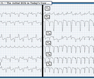
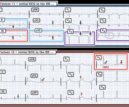
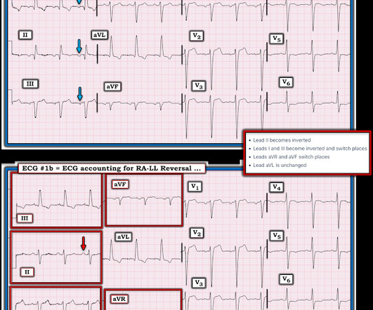
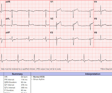
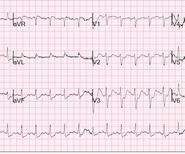
The ECG in Figure-1 — was obtained from a middle-aged man with palpitations and shortness of breath. How would YOU interpret the ECG in Figure-1 ? Figure-1: The initial ECG in today's case. ( To improve visualization — I've digitized the original ECG using PMcardio ). Figure-1: The initial ECG in today's case. (

EKG/ECG 442
article thumbnail

ECG Blog #436 — Bigeminy or Alternans?

Ken Grauer, MD

The ECG in Figure-1 — was obtained from an older man with known coronary disease. He developed cardiac arrest shortly after the ECG in Figure-1 was recorded. QUESTIONS: How would YOU interpret the ECG in Figure-1 ? QUESTIONS: How would YOU interpret the ECG in Figure-1 ? Figure-1: The initial ECG in today's case. (

EKG/ECG 401
Insiders

Sign Up for our Newsletter

This site is protected by reCAPTCHA and the Google Privacy Policy and Terms of Service apply.

Trending Sources

article thumbnail

ECG Blog #380 — What is "Swirl"?

Ken Grauer, MD

The ECG in Figure-1 — was obtained from an older woman with persistent CP ( C hest P ain ) over the previous day. Figure-1: The initial ECG in today's case. Voltage for LVH is satisfied — at least by Peguero Criteria ( Sum of deepest S in any chest lead + S in V4 ≥23 mm in a woman — as discussed in ECG Blog #73 ).

EKG/ECG 362
article thumbnail

ECG Blog #453 — Is this Wellens' Syndrome?

Ken Grauer, MD

The ECG in Figure-1 was obtained from a middle-aged man who presented with a 2 week history of progressively increasing CP ( C hest P ain ) with exertion. He had his most severe episode of CP the day before he was seen with this ECG. An initial Troponin drawn less than 1 hour after the ECG in Figure-1 was recorded was over 10,000.

EKG/ECG 236
article thumbnail

ECG Blog #375 — At Least 3 Major Findings.

Ken Grauer, MD

The ECG in Figure-1 was obtained from a 50-year old man — who presented to the ED ( E mergency D epartment ) with new-onset CP ( C hest P ain ). QUESTIONS: There are at least 3 principal findings on this ECG — some of which deal with a possible “culprit” artery and / or the location of whatever is going on.

EKG/ECG 195
article thumbnail

ECG Blog #387 — 2 Minutes Later.

Ken Grauer, MD

The ECG in Figure-1 was obtained from an elderly man with a history of coronary disease — who contacted EMS for "burning" chest discomfort that woke him at 3am. Some amount of time passed at home — during which his chest discomfort persisted The patient was hemodynamically stable at the time he was seen by EMS, when ECG #1 was recorded.

EKG/ECG 195
article thumbnail

Cath Lab occupied. Which patient should go now (or does only one need it? Or neither?)

Dr. Smith's ECG Blog

This was sent to me by an undergraduate name Hans Helseth, who is an EKG tech, but who is an expert OMI ECG reader. While an EKG and labs were being obtained on Patient 1, a second announcement came through for another prehospital “STEMI” activation on a 58 year old male ( Patient 2 ) with a history of some substance abuse.

EKG/ECG 119